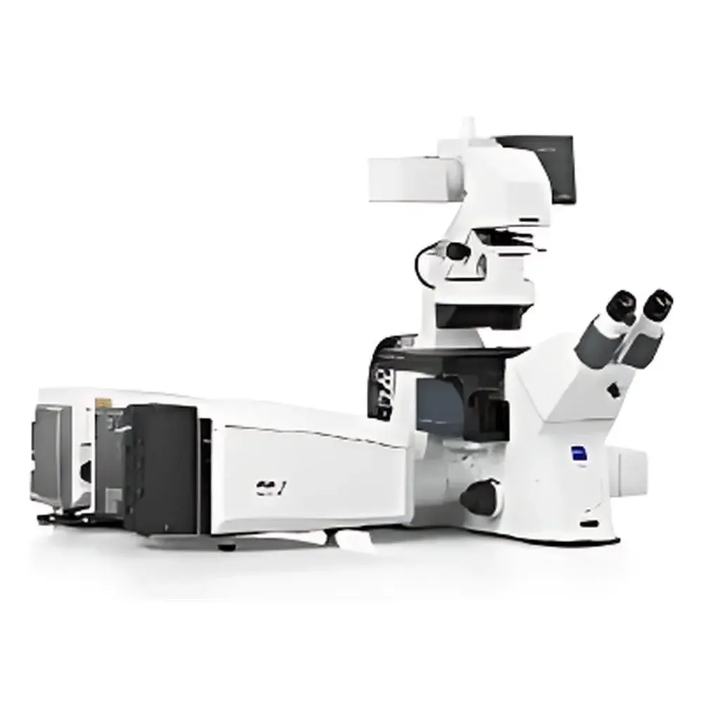
ZEISS LSM 990 Laser Scanning Confocal Microscope
| Brand | ZEISS |
|---|---|
| Origin | Germany |
| Instrument Type | Point-Scanning Confocal Microscope |
| Model | LSM 990 |
| Detector Technology | GaAsP Hybrid Detectors (HyD) & Airyscan 2 |
| Maximum Lateral Resolution | ≤80 nm (measured via FWHM of fluorescent beads) |
| Volumetric Imaging Speed | Up to 80 volumes/s |
| Excitation Sources | Integrated Multi-Line Lasers (405, 488, 561, 633 nm standard |
| Software Platform | ZEN Intellesis + ZEN Blue |
| Compliance | Designed for GLP/GMP-aligned workflows |
Overview
The ZEISS LSM 990 Laser Scanning Confocal Microscope is a high-performance, modular point-scanning confocal platform engineered for quantitative, multi-dimensional imaging in life science laboratories. Based on the principle of optical sectioning through spatially filtered detection of fluorescence emission, the LSM 990 eliminates out-of-focus light with precision pinhole optics and high-efficiency spectral beam splitting. Its core architecture integrates resonant and galvo scanning mechanisms, enabling both high-speed volumetric acquisition and diffraction-limited static imaging. The system supports simultaneous multi-channel detection across the visible to near-infrared spectrum (400–900 nm), with real-time spectral unmixing and linear unmixing algorithms embedded in ZEN software. Designed and manufactured in Oberkochen, Germany, the LSM 990 meets stringent ISO 9001 quality standards and is built for long-term stability under continuous operational loads typical in core imaging facilities and academic research labs.
Key Features
- Advanced detector suite: Dual GaAsP hybrid detectors (HyD) with >45% quantum efficiency and Airyscan 2 super-resolution module delivering up to 1.7× resolution gain over conventional confocal without compromising signal-to-noise ratio
- Flexible excitation architecture: Pre-aligned multi-line laser combiner with four standard solid-state lasers (405/488/561/633 nm); optional upgrade paths for UV (355 nm), white-light laser (WLL), or IR (780 nm) for two-photon compatibility
- High-throughput 4D imaging: Resonant scanner enables volumetric acquisition at up to 80 volumes per second—validated using live zebrafish embryonic heart imaging at 3 dpf
- Modular expansion capability: Native integration path for FLIM (fluorescence lifetime imaging), FCS (fluorescence correlation spectroscopy), FRAP (fluorescence recovery after photobleaching), and environmental control units (temperature, CO₂, humidity)
- ZEN Intellesis AI-assisted analysis: Built-in deep learning modules for segmentation, denoising, and object tracking—trained on annotated biological datasets and compliant with FAIR data principles
Sample Compatibility & Compliance
The LSM 990 accommodates a broad range of specimen formats including glass-bottom dishes (MatTek, WillCo), multi-well plates (96-/384-well), tissue sections (up to 1 mm thick with clearing), whole-mount embryos (zebrafish, Drosophila), and organoids cultured under physiological conditions. Its motorized stage and objective turrets support automated multi-position time-lapse experiments with sub-micron reproducibility. From a regulatory standpoint, the system is designed to support GLP-compliant documentation workflows. When configured with ZEN Blue v3.7+ and validated electronic signatures, it fulfills audit trail requirements per FDA 21 CFR Part 11. All calibration routines—including lateral resolution verification using NIST-traceable fluorescent microspheres (100 nm diameter)—are documented within the instrument’s internal logbook and exportable as PDF reports.
Software & Data Management
ZEN Blue serves as the primary acquisition and processing interface, offering intuitive wizard-driven experiment setup, hardware-synchronized metadata tagging (including laser power, dwell time, pinhole size, and objective ID), and hierarchical project organization. Raw data is stored in the open-format ZEN .czi file container, which embeds OME-TIFF-compatible metadata and supports direct import into ImageJ/Fiji, Python (via czifile and aicsimageio libraries), and MATLAB. For enterprise environments, optional ZEN Connect enables centralized license management, remote instrument monitoring, and secure DICOM export for translational imaging pipelines. All software updates undergo ISO/IEC 17025-aligned validation protocols prior to release.
Applications
The LSM 990 is routinely deployed in studies requiring quantitative subcellular localization, dynamic protein trafficking, and multiplexed structural phenotyping. Representative use cases include: 3D reconstruction of synaptic architecture in primary neuronal cultures; longitudinal tracking of mitochondrial fission/fusion events in live iPSC-derived cardiomyocytes; spectral separation of overlapping fluorophores in multicolor plant root tip imaging; quantification of nuclear translocation kinetics following cytokine stimulation; and correlative analysis of vascular perfusion dynamics and endothelial junction integrity in aged murine brain slices. Its compatibility with expansion modules makes it suitable for method development in super-resolution biophysics, including single-molecule counting and diffusion coefficient mapping via raster image correlation spectroscopy (RICS).
FAQ
What is the minimum achievable lateral resolution under Airyscan 2 mode?
Measured using 100 nm fluorescent beads and deconvolved with Joint-DCV algorithm, the system achieves ≤80 nm full-width half-maximum (FWHM) in xy-plane at 63×/1.4 NA oil immersion.
Can the LSM 990 be integrated into an existing two-photon microscope setup?
Yes—the system supports external laser input via fiber-coupled ports and synchronization signals for hybrid modalities, provided the host platform provides TTL-triggered scan mirror control and galvo synchronization.
Is ZEN software compatible with Linux-based HPC clusters for batch processing?
ZEN Blue runs natively on Windows 10/11; however, .czi files can be processed on Linux clusters using open-source tools such as aicsimageio, napari, or custom Python pipelines—no proprietary runtime dependency required.
Does the system support automated focus maintenance during long-term live-cell imaging?
Yes—hardware autofocus (Definite Focus 2) is standard and operates independently of the scanning process, maintaining focus stability within ±50 nm over 24-hour acquisitions.
How is calibration traceability maintained for quantitative intensity measurements?
Each detector channel is calibrated annually using ZEISS-certified reference standards (fluorescent microspheres, neutral density filters), with calibration certificates linked to instrument logs and exportable upon request.


