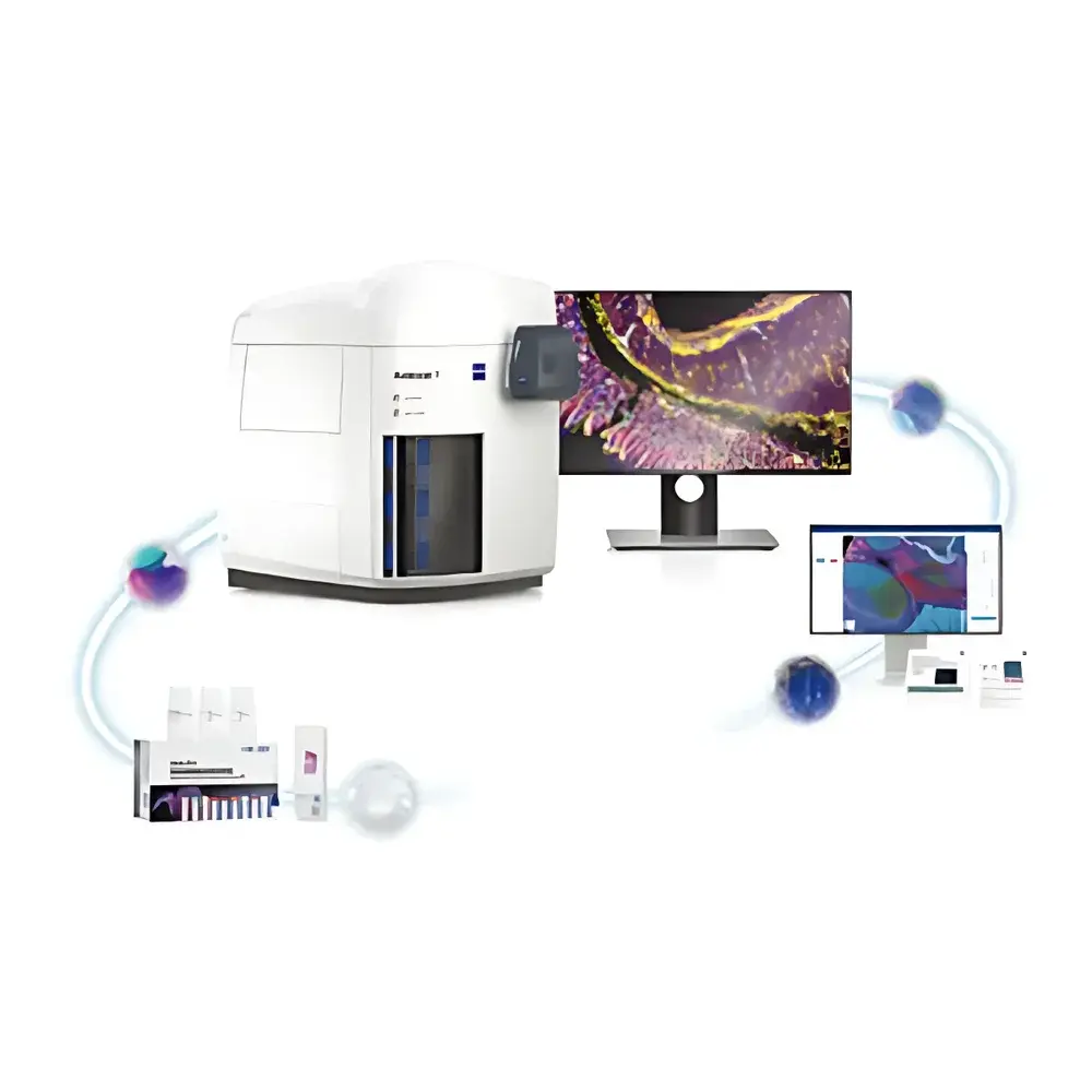
ZEISS RIMA Spatial Biology Solution
| Brand | ZEISS |
|---|---|
| Origin | Shanghai, China |
| Manufacturer Type | Authorized Distributor |
| Product Origin | Domestic (China) |
| Model | RIMA Spatial Biology Solution |
| Pricing | Upon Request |
Overview
The ZEISS RIMA Spatial Biology Solution is an integrated, end-to-end platform engineered for high-plex, spatially resolved multiplex immunofluorescence (mIF) imaging and quantitative analysis. Built upon ZEISS’s proven optical precision and modular microscope architecture—typically leveraging the ZEISS Axio Imager or ZEISS LSM 900 platforms—the RIMA system implements iterative immunolabeling, automated cyclic staining, and spectral unmixing–enabled image acquisition to resolve ≥6–12 biomarkers per tissue section with subcellular spatial fidelity. Its core measurement principle relies on sequential antibody stripping and re-probing combined with high-sensitivity sCMOS or GaAsP detector-based fluorescence imaging, enabling accurate co-localization mapping and neighborhood-based cellular phenotyping within native tissue architecture. Designed for translational research laboratories operating under GLP-aligned workflows, the solution bridges discovery-stage spatial biology with preclinical biomarker validation and clinical assay development.
Key Features
- Fully automated, software-guided cyclic immunostaining workflow—minimizing manual intervention and inter-operator variability across serial labeling rounds.
- Integrated hardware synchronization between motorized stage, filter wheels, objective turrets, and LED/laser excitation sources to ensure pixel-registered image stacks across cycles.
- Robust signal preservation through optimized antigen retrieval, low-background blocking, and gentle antibody elution chemistry—validated for FFPE and frozen sections up to 10 µm thickness.
- Real-time quality control metrics embedded in acquisition software—including focus stability tracking, intensity drift correction, and cycle-to-cycle registration error quantification (RMS < 0.3 µm).
- Modular scalability: compatible with ZEISS ZEN Blue/Black acquisition environments and expandable to include AI-assisted segmentation (e.g., Deep Learning-based nuclear/cytoplasmic boundary detection) via optional plugins.
Sample Compatibility & Compliance
The RIMA platform supports standard histological formats including formalin-fixed paraffin-embedded (FFPE) tissue sections (4–10 µm), cryosections, and cytospin preparations. It complies with ISO 13485–aligned manufacturing controls (as applied by ZEISS-certified integration partners in Shanghai), and data handling workflows support audit-trail generation per FDA 21 CFR Part 11 requirements when deployed with ZEN Connect and ZEISS Lab Archive modules. All staining protocols are developed in alignment with CAP/CLIA pre-analytical guidelines and referenced against ASTM E3107–20 standards for multiplex tissue imaging reproducibility.
Software & Data Management
Acquisition, registration, spectral unmixing, and cell segmentation are orchestrated through ZEISS ZEN software (v3.6+), with native support for OME-TIFF export and interoperability with QuPath, HALO, and Visiopharm via standardized .qptiff and .ndpi wrappers. The RIMA-specific workflow manager enforces version-controlled protocol templates, automatic metadata tagging (including antibody clone, lot number, dilution, and incubation time), and encrypted project archiving. Raw image datasets are structured according to the MIAME-compliant spatial omics metadata schema, facilitating downstream integration into federated analysis platforms such as SpatialDB or the Human Tumor Atlas Network (HTAN) submission pipeline.
Applications
- Immuno-oncology biomarker discovery—quantifying PD-1/PD-L1 co-expression patterns, T-cell exclusion metrics, and tertiary lymphoid structure (TLS) density in NSCLC and melanoma cohorts.
- Spatial characterization of tumor-immune microenvironments in response to checkpoint inhibitor therapy or adoptive cell transfer regimens.
- Development of spatially informed diagnostic classifiers for companion diagnostics, aligned with IVD regulatory pathways (e.g., CE-IVDR Annex II Class C criteria).
- High-throughput validation of single-cell RNA-seq-derived cell type signatures in situ—enabling cross-platform concordance assessment between transcriptomic and proteomic spatial layers.
FAQ
What tissue types and thicknesses are validated for use with the RIMA workflow?
FFPE sections from 4–10 µm and OCT-embedded frozen sections up to 8 µm have been experimentally validated using ZEISS-recommended antigen retrieval and staining protocols.
Does the RIMA solution support integration with third-party analysis tools like QuPath or HALO?
Yes—ZEN software exports registered, unmixed TIFF stacks with embedded coordinate transforms, enabling direct import into QuPath (v0.4.0+) and HALO (v3.2+) with preserved spatial topology and marker channel assignment.
Is the system compliant with regulatory documentation requirements for clinical assay development?
When configured with ZEISS Lab Archive and ZEN Connect audit modules, the system supports electronic signatures, change logs, and full traceability of acquisition parameters—meeting baseline expectations for GLP and early-phase IVD assay validation.
How is staining reproducibility ensured across multiple instruments or sites?
RIMA leverages ZEISS’s centralized protocol repository and hardware calibration routines (e.g., LED intensity normalization, auto-focus reference tile mapping), enabling cross-site harmonization verified through multi-center ring trials per ISO/IEC 17043 guidelines.


