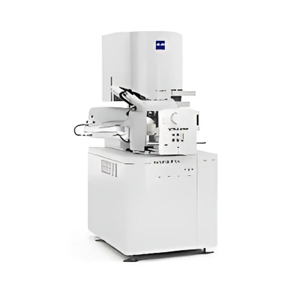
ZEISS Volutome In-Situ High-Throughput Microtome for FE-SEM
| Brand | ZEISS |
|---|---|
| Origin | Germany |
| Model | Volutome |
| Application | Integrated in-chamber ultramicrotome for serial block-face imaging in field-emission scanning electron microscopy (FE-SEM) |
| Detector | Volume BSD (Backscattered Electron Detector optimized for volumetric surface imaging) |
| Charge Compensation | Patented localized charge compensation system |
| Automation | Unattended operation up to 72 hours |
| Workflow Integration | End-to-end solution from sectioning and imaging to 3D reconstruction and visualization |
Overview
The ZEISS Volutome is an in-chamber, high-throughput ultramicrotome engineered exclusively for integration with ZEISS field-emission scanning electron microscopes (FE-SEM), including the GeminiSEM series. It enables automated serial block-face sectioning and imaging of resin-embedded biological specimens—such as brain tissue, plant organs, or cultured cells—within the SEM vacuum chamber. Unlike conventional ex-situ microtomes, the Volutome eliminates manual handling, stage repositioning, and environmental exposure between cuts, thereby preserving structural integrity and enabling true volumetric imaging at nanoscale resolution. Its operational principle relies on precise piezo-driven diamond-knife advancement synchronized with high-speed backscattered electron (BSE) detection using the proprietary Volume BSD detector. This architecture supports acquisition of large-volume 3D datasets with sub-10 nm isotropic voxel resolution, while maintaining specimen stability under low-accelerating-voltage conditions (≤1.5 kV) to minimize beam-induced damage and charging artifacts.
Key Features
- In-chamber integration: Fully embedded within ZEISS FE-SEM systems (e.g., GeminiSEM 360/460), eliminating air exposure and mechanical misalignment during serial sectioning.
- Volume BSD detector: A high-efficiency, high-dynamic-range backscattered electron detector specifically designed for volumetric surface contrast, delivering superior signal-to-noise ratio for resin-embedded biological sections without metal coating.
- Patented localized charge compensation system: Actively neutralizes surface charge accumulation on insulating samples (e.g., epoxy-embedded tissues) during low-kV imaging, ensuring consistent contrast and dimensional fidelity across extended acquisition sessions.
- 72+ hour unattended operation: Robust thermal and mechanical stability, coupled with real-time knife wear monitoring and adaptive cutting algorithms, enables fully autonomous data collection over multiple days.
- Synchronized imaging and preprocessing: On-the-fly image denoising, contrast normalization, and drift correction occur during acquisition—reducing post-processing latency and accelerating time-to-3D-reconstruction.
- Modular workflow architecture: Hardware control, acquisition scheduling, and 3D rendering are unified under ZEISS SmartSEM and arivis Vision4D software environments, supporting reproducible, auditable, and GLP-compliant experimental protocols.
Sample Compatibility & Compliance
The Volutome accommodates standard 3 mm × 3 mm × 1 mm resin blocks (e.g., Durcupan, Epon, LR White), compatible with conventional chemical fixation, osmium tetroxide post-fixation, and en bloc staining protocols. It supports both hard and semi-hard embedding media, including acrylics used in plant ultrastructure studies. All hardware and software components comply with CE marking requirements and meet electromagnetic compatibility (EMC) standards per EN 61326-1. The arivis Vision4D platform supports audit trails, user access controls, and electronic signatures in accordance with FDA 21 CFR Part 11 for regulated preclinical research environments. Data export formats (e.g., TIFF stacks, HDF5, N5) ensure interoperability with open-source reconstruction tools such as Ilastik, Fiji, and CATMAID.
Software & Data Management
Acquisition and reconstruction are managed through two tightly coupled software layers: ZEISS SmartSEM for instrument control and real-time parameter adjustment (knife advance step, dwell time, pixel size, frame averaging), and arivis Vision4D for automated stitching, alignment, segmentation, and volume rendering. Vision4D implements GPU-accelerated registration algorithms capable of correcting nonlinear distortions arising from knife chatter or thermal drift. Raw image metadata—including acceleration voltage, working distance, detector gain, and timestamped cut thickness—is embedded directly into TIFF headers and preserved throughout the processing pipeline. Batch processing scripts support standardized pipelines compliant with MIAP (Minimum Information About a 3D Imaging Experiment) guidelines, facilitating data deposition in public repositories such as EMPIAR or BioImage Archive.
Applications
- Neuroscience: Connectomic mapping of mammalian brain tissue at synaptic resolution; quantification of mitochondrial morphology, synaptic vesicle density, and myelin periodicity in models of neurodegeneration, aging, or memory consolidation.
- Cell biology: Statistical analysis of organelle number, size distribution, and spatial organization across cell populations under physiological vs. pathological states (e.g., autophagy induction, ER stress response).
- Plant science: Subcellular phenotyping of chloroplast ultrastructure, plasmodesmata distribution, and cell wall modifications in response to biotic/abiotic stress or genetic perturbation.
- Developmental biology: 3D reconstruction of embryonic tissue morphogenesis at organ-level scale, combining serial sectioning with immunogold labeling for correlative ultrastructural localization.
- Pharmacology & toxicology: Assessment of drug-induced ultrastructural changes in liver sinusoids, renal tubules, or cardiac myofibrils following chronic dosing regimens.
FAQ
Is the Volutome compatible with non-ZEISS SEM platforms?
No—the Volutome is a purpose-built, mechanically and electronically integrated subsystem requiring native communication with ZEISS GeminiSEM control firmware and vacuum interlocks.
What is the minimum achievable section thickness?
Typical stable operation ranges from 10 nm to 100 nm, depending on sample hardness and knife geometry; 30 nm is routinely used for neuronal connectomics.
Can stained or unstained samples be imaged?
Both are supported; heavy-metal-stained samples yield higher BSE contrast, but unstained sections can be imaged effectively using the Volume BSD detector’s enhanced sensitivity at low kV.
How is knife wear monitored during long acquisitions?
Real-time BSE signal amplitude tracking and edge sharpness analysis trigger automatic knife repositioning or user alerts when cut quality degrades beyond defined thresholds.
Does the system support correlative light and electron microscopy (CLEM)?
Yes—integrated fiducial marker recognition and coordinate mapping enable precise registration of fluorescence microscopy data acquired prior to SEM mounting.


