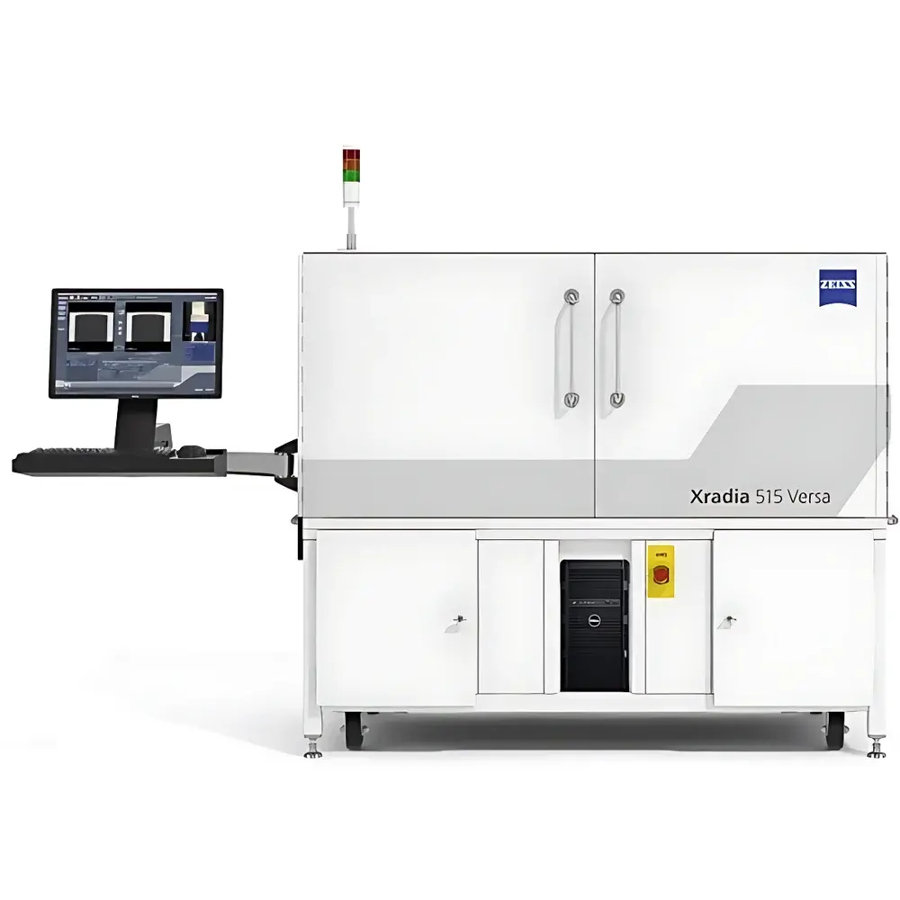
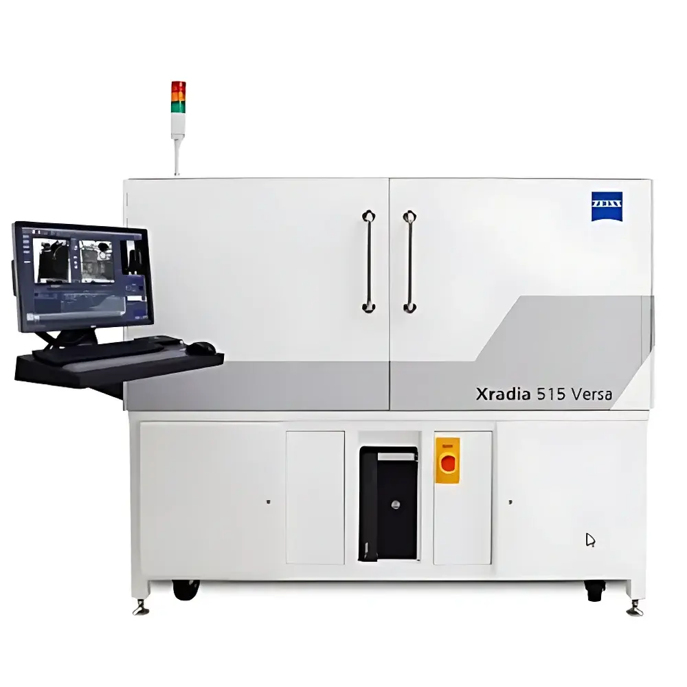
ZEISS Xradia 515 Versa X-ray Microscope
| Brand | ZEISS |
|---|---|
| Origin | Germany |
| Model | Xradia 515 Versa |
| Spatial Resolution | ≤500 nm (true native resolution) |
| Imaging Modes | Absorption Contrast, Propagation-Based Phase Contrast |
| Software Platform | Scout-and-Scan Control System with ART 3.0 Advanced Reconstruction Toolbox |
| Compatibility | In Situ Mechanical & Thermal Stages, Automated Sample Handling, Flat-Panel Detector Options |
| Regulatory Compliance | Designed for GLP/GMP-aligned workflows |
Overview
The ZEISS Xradia 515 Versa is a high-performance laboratory-based 3D X-ray microscope engineered for non-destructive, multi-scale tomographic imaging across diverse scientific and industrial domains. Unlike conventional micro-CT systems constrained by geometric magnification limits or source-detector distance trade-offs, the Xradia 515 Versa employs a proprietary synchrotron-inspired architecture—featuring a high-brightness rotating anode X-ray source, precision reflective optics (capillary condenser and zone plate objectives), and a high-dynamic-range CMOS detector—to deliver true spatial resolution down to 500 nm at working distances exceeding 10 mm. This optical design enables high-fidelity phase contrast and absorption contrast imaging without requiring sample sectioning, metal coating, or vacuum environments. The system operates in the 5–90 keV energy range, supporting both low-Z biological tissues and high-Z metallic or ceramic specimens. Its mechanical stability, thermal drift compensation, and vibration-isolated optical path ensure measurement repeatability essential for longitudinal studies and quantitative morphometric analysis.
Key Features
- True 500 nm spatial resolution maintained across large field-of-view (up to 1.2 mm × 1.2 mm) and extended working distances (≥10 mm), enabling high-resolution imaging of intact, unmodified samples ranging from millimeter-scale battery electrodes to centimeter-scale rock cores.
- Dual-contrast capability: Simultaneous acquisition and separation of absorption and propagation-based phase contrast signals via edge-enhancement algorithms—critical for visualizing low-density interfaces in polymers, soft tissues, or porous media.
- Intelligent collision avoidance system: Real-time motorized stage position monitoring combined with 3D sensor feedback prevents physical contact between objective optics and sample during automated scan setup and multi-position acquisition.
- Modular expandability: Native support for in situ mechanical testing stages (tensile/compression), environmental chambers (−20 °C to +200 °C), 4D time-resolved tomography modules, and robotic sample changers—all integrated via standardized ZEISS hardware abstraction layer (HAL).
- Optimized reconstruction pipeline: Fully compatible with ART 3.0 Advanced Reconstruction Toolbox, incorporating iterative regularization, deep learning–assisted denoising, and GPU-accelerated FDK and SART solvers for artifact suppression and quantitative density calibration.
Sample Compatibility & Compliance
The Xradia 515 Versa accommodates heterogeneous sample geometries—including irregular, fragile, hydrated, or electrically conductive specimens—without preparation artifacts. It complies with ASTM E1441 (Standard Guide for Computed Tomography), ISO/IEC 17025 requirements for measurement uncertainty estimation, and supports traceable calibration using NIST-traceable step wedges and resolution test patterns. When configured with validated software modules and documented SOPs, the system meets analytical data integrity expectations under FDA 21 CFR Part 11 and EU Annex 11 for regulated research environments. Full metadata embedding (source parameters, geometry, reconstruction settings) ensures reproducibility and audit readiness.
Software & Data Management
Acquisition, reconstruction, and analysis are unified under the Scout-and-Scan Control System—a deterministic, scriptable platform supporting Python API integration. Raw projections are stored in HDF5 format with embedded provenance metadata. ART 3.0 provides batch processing, region-of-interest masking, porosity quantification (per ASTM D7908), fiber orientation analysis (ISO 13067), and segmentation-ready outputs compatible with Avizo, Dragonfly, and MATLAB. All user actions—including parameter changes, reconstruction iterations, and annotation events—are logged with timestamps and operator IDs, satisfying GLP documentation requirements.
Applications
- Materials Science: Quantitative 3D characterization of pore networks in additively manufactured alloys, crack propagation in thermal barrier coatings, and interfacial delamination in composite laminates.
- Life Sciences: Microarchitecture analysis of trabecular bone, vascular network mapping in decalcified tissue, and developmental morphology in whole-organism embryonic models.
- Earth Sciences: Pore-throat connectivity modeling in reservoir rocks, fossil internal morphology reconstruction, and fluid flow simulation input generation.
- Electronics & Semiconductors: Void detection in solder joints, through-silicon via (TSV) integrity assessment, and package-level failure root cause analysis without destructive cross-sectioning.
- In Situ & 4D Studies: Real-time monitoring of electrochemical processes in operando battery cells, creep deformation in high-temperature superalloys, and hydration-driven swelling in cementitious materials.
FAQ
What distinguishes the Xradia 515 Versa from conventional micro-CT systems?
It utilizes reflective X-ray optics rather than geometric magnification alone, enabling true sub-500 nm resolution without sacrificing field of view or working distance.
Can the system perform quantitative density measurements?
Yes—when calibrated with reference phantoms and operated under stabilized beam conditions, it supports linear attenuation coefficient mapping with ±3% relative uncertainty across a dynamic range of 0.01–10 cm⁻¹.
Is remote operation supported?
The Scout-and-Scan platform includes secure TLS-encrypted remote desktop access and RESTful API endpoints for integration into automated lab infrastructure.
Does ZEISS provide application-specific method development support?
Yes—ZEISS Application Scientists collaborate on SOP development, including reconstruction parameter optimization, segmentation workflow validation, and uncertainty budgeting per ISO/IEC 17025.
What maintenance intervals are recommended for long-term stability?
Source anode refurbishment every 18–24 months (usage-dependent); annual optical alignment verification and detector gain calibration are advised per ZEISS Service Protocol SP-XRM-001.


