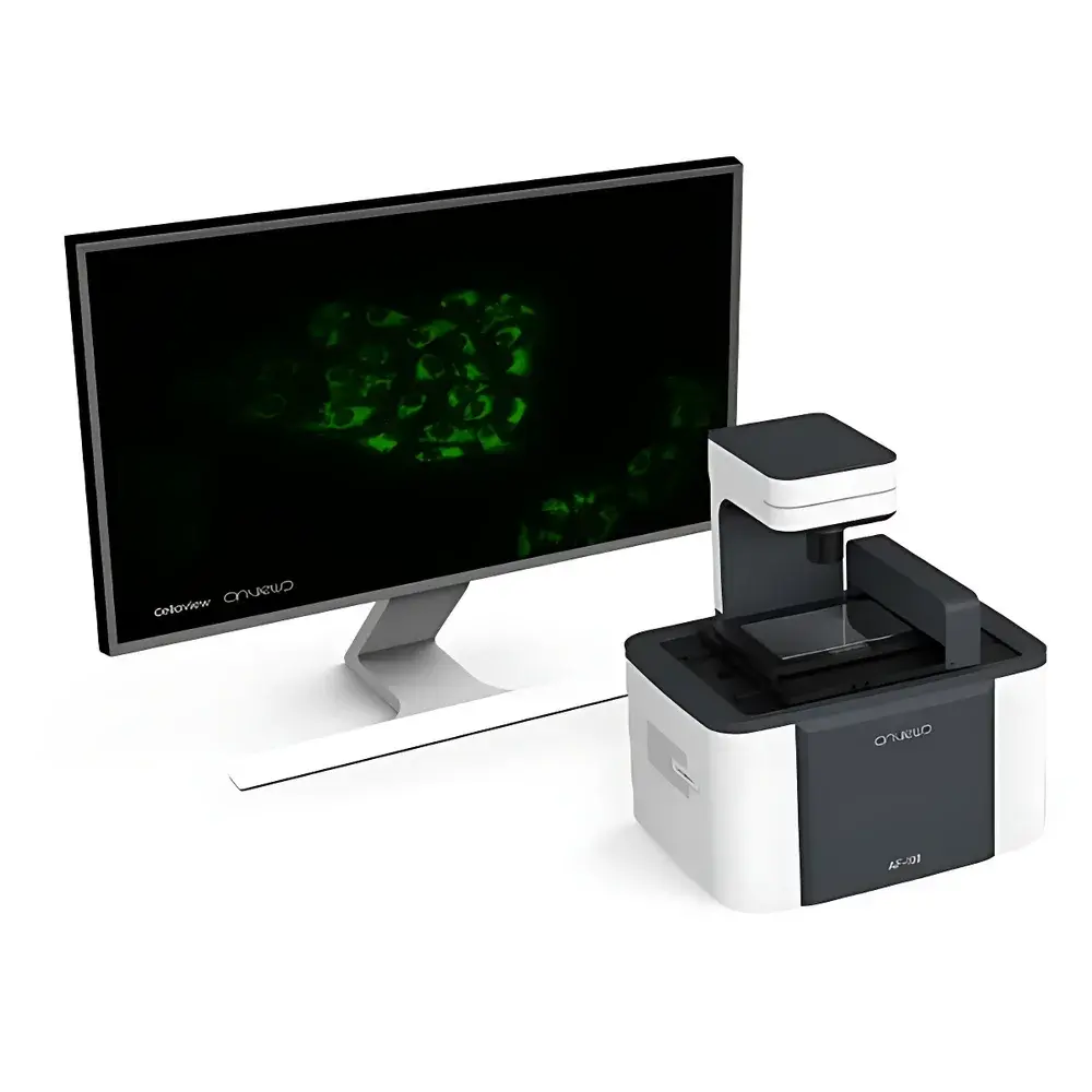
Aure AF100 Live-Cell Imaging and Analysis System
| Brand | Aure |
|---|---|
| Model | AF100 |
| Origin | Jiangsu, China |
| Instrument Type | Automated Time-Lapse Microscopy System for Long-Term Culture Monitoring |
| Optical Resolution | 5 MP Monochrome CMOS Sensor |
| Compatible Well Plates | 6–384-well |
| Objective Lenses Supported | 4×, 10×, 20× (Standard, UP, and LWD) |
| Imaging Mode | Brightfield Only |
| Maximum Scan Speed | Full 96-well Plate in ≤4 min |
| Environmental Integration | Designed for In-CO₂ Incubator Deployment (Temperature/Humidity/CO₂ Stable Operation) |
| Software | Integrated Time-Lapse Acquisition, Image Stitching, Video Synthesis, and Multi-Point Retrospective Analysis Engine |
Overview
The Aure AF100 Live-Cell Imaging and Analysis System is an engineered solution for non-invasive, long-term quantitative monitoring of adherent and suspension mammalian cells under physiologically relevant culture conditions. Unlike conventional benchtop microscopes requiring sample removal from controlled environments, the AF100 integrates directly into standard CO₂ incubators—enabling continuous, label-free brightfield observation without thermal or gas-phase perturbation. Its operational principle relies on automated time-lapse microscopy: a precision X-Y-Z-θ motorized stage positions the specimen relative to a fixed optical path, while a high-sensitivity 5-megapixel monochrome CMOS sensor captures sequential images at user-defined intervals. The system’s optical architecture incorporates aberration-corrected illumination pathways specifically tuned to mitigate meniscus-induced artifacts (e.g., “bull’s-eye” and “crescent” distortions) commonly observed in multi-well plate imaging—ensuring uniform intensity distribution across 96- and 384-well formats. This design supports longitudinal tracking of confluence dynamics, morphological transitions, mitotic events, and cytotoxic responses with temporal resolution down to 1 minute.
Key Features
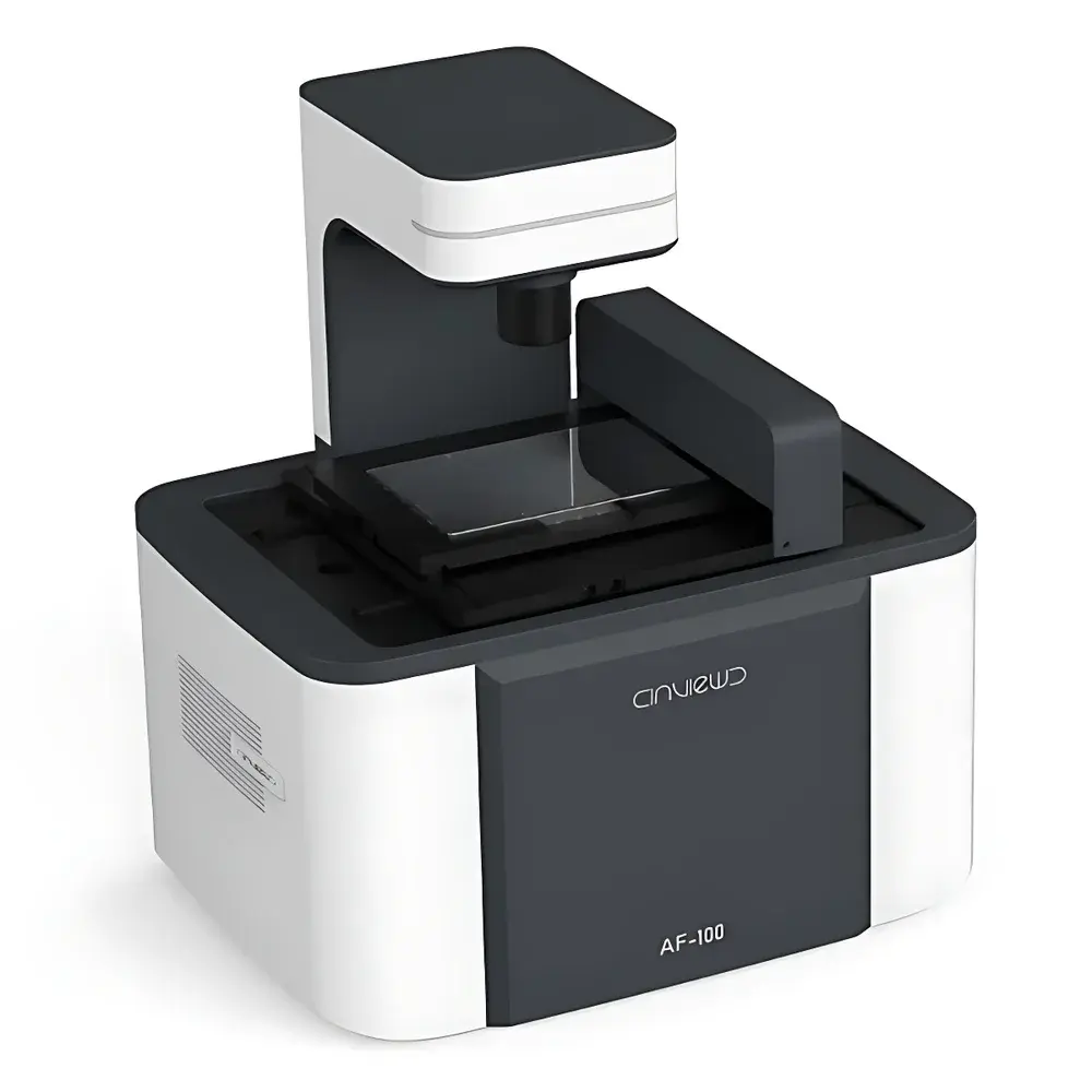
- Incubator-integrated form factor: Compact footprint (W × D × H < 200 × 200 × 180 mm) enables deployment inside standard 180–220 L CO₂ incubators without obstructing airflow or humidity distribution.
- Multi-objective compatibility: Accepts standard 4×, 10×, and 20× objectives—including ultra-plan (UP) and long-working-distance (LWD) variants—to accommodate varied vessel heights (e.g., Petri dishes, microfluidic chips, and deep-well plates).
- Automated multi-field acquisition: Four-axis (X-Y-Z-θ) motion control enables programmable scanning across up to 16 fields per well, with dynamic focus adjustment per position to compensate for well-bottom curvature.
- High-throughput imaging cycle: Full 96-well plate acquisition completed in ≤4 minutes using optimized exposure and stage motion algorithms—minimizing cumulative phototoxicity and thermal drift.
- Embedded video synthesis engine: Raw image sequences are automatically compiled into time-stamped AVI or MP4 files with embedded metadata (timestamp, well ID, objective used, focus offset), eliminating post-acquisition scripting requirements.
Sample Compatibility & Compliance
The AF100 supports routine monitoring of primary human fibroblasts, iPSC-derived lineages, HEK293, HeLa, CHO-K1, and other adherent or low-density suspension cultures grown in standard tissue-culture-treated polystyrene or glass-bottom multi-well plates. It complies with ISO 13485-aligned manufacturing controls as applied to laboratory instrumentation, and its software architecture supports audit-trail generation per GLP and GMP documentation standards. While not FDA 21 CFR Part 11–validated out-of-the-box, the system’s logging framework records all acquisition parameters, user actions, and timestamped image metadata—facilitating internal validation protocols required for regulated bioprocess development or preclinical assay qualification.
Software & Data Management
The AF100 Control Suite provides a Windows-based interface for experiment scheduling, hardware calibration, and real-time preview. Acquired data are stored in hierarchical folder structures compliant with MIAME and MIAPE metadata conventions. Built-in analysis modules include confluence quantification (threshold-based pixel classification), phase-contrast contrast enhancement (non-linear histogram stretching), and region-of-interest (ROI) time-series export in CSV/TXT format for downstream statistical modeling in MATLAB, Python (NumPy/Pandas), or GraphPad Prism. All image files retain EXIF-compatible tags containing exposure time, gain, lens focal length, and environmental log snippets (if connected to optional incubator telemetry interfaces).
Applications
- Longitudinal assessment of cell proliferation kinetics during monoclonal antibody production process development.
- Real-time detection of morphological hallmarks associated with apoptosis or senescence in CRISPR-edited isogenic cell lines.
- Optimization of feeder-layer-dependent stem cell maintenance protocols via daily confluence mapping across 384-well arrays.
- Quantitative comparison of cytotoxicity onset across dose gradients in 96-well drug screening assays—without endpoint staining or trypsinization.
- Validation of bioreactor inoculum quality by correlating early adhesion efficiency (T = 2–6 h) with subsequent expansion yield.
FAQ
Can the AF100 be used outside of a CO₂ incubator?
Yes—though its thermal management and vibration-damping chassis are optimized for incubator mounting, it operates stably on benchtops with ambient temperature control (18–28°C) and stable power supply.
Does the system support fluorescence imaging?
No—the AF100 is a dedicated brightfield platform; fluorescence capability requires external light sources, filter cubes, and emission optics not included in the base configuration.
Is remote access supported for off-site monitoring?
Yes—via secure RDP or VNC over local network; no cloud hosting or internet-exposed services are enabled by default to maintain data sovereignty.
What file formats are generated during acquisition?
TIFF (uncompressed, 16-bit), JPEG (lossless compression), and MP4 (H.264-encoded video); metadata embedded in XMP sidecar files.
How is focus stability maintained across multi-day acquisitions?
Through periodic auto-focus routines triggered by user-defined intervals or confluence thresholds, using contrast gradient maximization algorithms applied to central ROI subframes.


