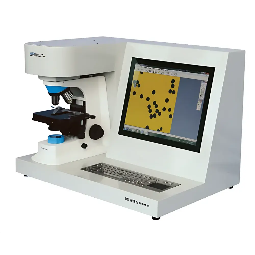
Yidian Woguang WJL-708 Intelligent Particle Image Analyzer (with Domestic Microscope)
| Brand | Yidian Woguang (YDWG) |
|---|---|
| Origin | Shanghai, China |
| Model | WJL-708 |
| Dispersion Method | Dry Dispersion |
| Instrument Type | Laboratory Particle Image Analyzer |
| Measurement Range | 0.1–3000 µm |
| Maximum Magnification | 8000× |
| Spatial Resolution | 0.1 µm/pixel |
| Repeatability | ≤ ±1% |
| Automatic Segmentation Speed | ≤ 1 s per image |
| CCD Camera | 5 MP |
| Data Storage | Integrated 128 GB SSD + 15″ Display |
| Interface | USB / Ethernet / Wi-Fi |
| Power Supply | AC 220 V ±10%, 50 Hz, 200 W |
| Dimensions | 625 × 420 × 435 mm |
| Net Weight | 25 kg |
Overview
The Yidian Woguang WJL-708 Intelligent Particle Image Analyzer is a laboratory-grade particle characterization system that implements digital image analysis to quantify morphological and dimensional properties of solid particulates. Unlike laser diffraction or dynamic light scattering instruments, the WJL-708 employs high-magnification optical microscopy coupled with a calibrated 5-megapixel CCD imaging subsystem and deterministic image segmentation algorithms to deliver direct, two-dimensional geometric measurements—including equivalent circular diameter, aspect ratio, convexity, roundness, perimeter, projected area, and Feret diameters. This approach provides traceable, visually verifiable data essential for R&D validation, QC release testing, and regulatory documentation where particle shape—not just size—impacts functional performance (e.g., flowability, dissolution rate, packing density, or aerosol dispersion efficiency). The system operates at up to 8000× total magnification with a spatial resolution of 0.1 µm/pixel, enabling reliable analysis across a broad dynamic range from submicron agglomerates to coarse granules (0.1–3000 µm). Its dry dispersion module ensures representative sampling of free-flowing powders without liquid-mediated artifacts.
Key Features
- Integrated optical platform featuring a domestically manufactured high-stability microscope with precision mechanical stage and LED Köhler illumination for uniform, glare-free field illumination.
- Dedicated 5 MP progressive-scan CCD camera with low-noise CMOS sensor and real-time digitization at 30 fps, optimized for high-fidelity grayscale capture under variable contrast/brightness conditions.
- Proprietary image processing software with over 30 algorithmic modules—including adaptive thresholding, morphological filtering, multi-scale edge detection, local contrast enhancement, and intelligent auto-segmentation completing in ≤1 second per frame.
- Comprehensive geometric parameter library: computes >25 shape descriptors (e.g., circularity, solidity, elongation, convex hull ratio) and size metrics (area-equivalent diameter, Feret X/Y, Martin length) on a per-particle basis.
- Statistical engine supporting linear and logarithmic binning, cumulative/differential distribution plots, and customizable export formats (CSV, PNG, PDF) compliant with internal SOPs and external audit requirements.
- Self-contained workstation architecture: embedded industrial PC with 128 GB SSD, 15″ touchscreen display, and native support for USB, Gigabit Ethernet, and IEEE 802.11n wireless connectivity.
Sample Compatibility & Compliance
The WJL-708 accommodates dry, non-adhesive powders and granular solids with minimal pretreatment—ideal for pharmaceutical excipients, catalyst supports, ceramic precursors, metal AM feedstocks, and mineral aggregates. Sample loading is performed manually via standardized sample holders compatible with ISO 13322-1:2014 (Particle size analysis — Image analysis methods — Part 1: Static image analysis). While the instrument itself does not carry CE/UKCA marking, its measurement methodology aligns with ASTM E2457–22 (Standard Practice for Determining Particle Size Distribution of Powders Using Static Image Analysis) and supports GLP-compliant workflows through configurable user access levels, electronic signature prompts, and full audit trail logging (including operator ID, timestamp, parameter settings, and raw image metadata). System calibration is traceable to NIST-traceable stage micrometers; routine verification uses certified reference microspheres (e.g., Duke Scientific 6.0 µm PS-L).
Software & Data Management
The embedded analysis software runs on a locked-down Windows IoT Enterprise OS, preconfigured with validated firmware and no third-party bloatware. All image acquisition, preprocessing, segmentation, measurement, and reporting occur within a single unified interface. Raw images are stored with EXIF metadata (exposure time, gain, lens ID, calibration timestamp), while processed results include full statistical summaries (mean, D10/D50/D90, span, skewness) and scatter plots correlating shape vs. size parameters. Data exports comply with 21 CFR Part 11 requirements when configured with optional PKI-based digital signatures and role-based permissions (Administrator, Analyst, Reviewer). Backups can be scheduled to network shares or external drives via SMB or SFTP protocols; version history is retained for ≥90 days by default.
Applications
- Pharmaceutical development: Quantifying crystal habit changes during polymorph screening or milling process optimization.
- Advanced ceramics: Correlating green-body density with particle angularity and packing efficiency.
- Battery materials: Assessing secondary particle integrity and primary agglomerate morphology in cathode/anode powders.
- Food science: Monitoring starch granule swelling kinetics or fat crystal network evolution under thermal cycling.
- Quality control labs: Validating batch-to-batch consistency of spray-dried lactose or milled API blends against approved reference images.
- Academic research: Supporting publications requiring morphologically resolved particle statistics rather than ensemble-averaged diffraction profiles.
FAQ
Does the WJL-708 require wet dispersion or liquid suspension?
No—it is configured exclusively for dry dispersion using gravity-fed or vibratory sample introduction. Liquid-based analysis is not supported.
Can the system analyze transparent or low-contrast particles?
Yes—through adjustable LED illumination intensity, polarization filters, and multi-frame stacking algorithms that enhance edge definition without staining or coating.
Is third-party software integration possible?
Raw image streams (BMP/TIFF) and structured CSV outputs are fully accessible; however, the proprietary analysis engine does not expose APIs or DLL interfaces for external control.
What maintenance is required for long-term calibration stability?
Annual verification using traceable microsphere standards is recommended; optical alignment checks should be performed quarterly if used in environments with temperature fluctuations >±2°C/hour.
How is data integrity ensured during power interruption?
The embedded SSD includes power-loss protection circuitry; unsaved acquisitions are automatically cached and recovered upon reboot without file corruption.


