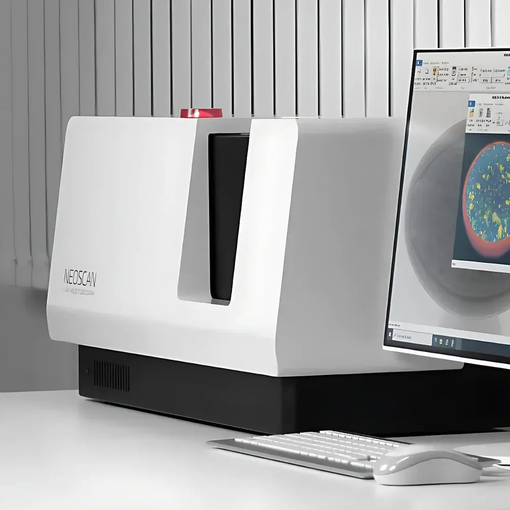
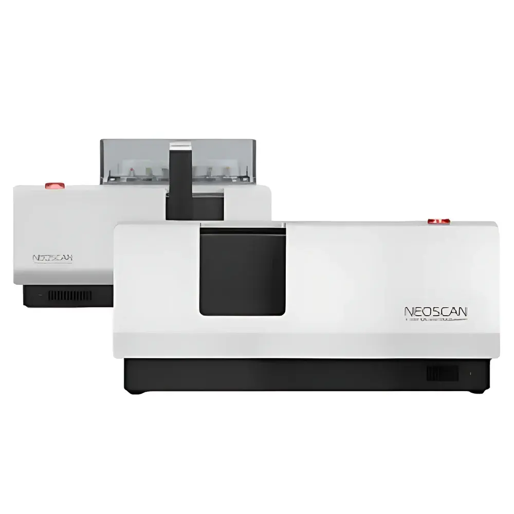
Neoscan N60 Compact Benchtop Micro-CT System
| Brand | Neoscan |
|---|---|
| Origin | Belgium |
| Model | N60 |
| X-ray Source Energy | 100 kV |
| Detector Type | Flat-panel Detector |
| Scan Mode | Rotation-Only (RO) |
| Spatial Resolution | 8 µm |
| Measurement Field of View | 36 mm × 60 mm |
| Sample Weight Capacity | 20 kg |
| System Dimensions | 800 mm × 310 mm × 375 mm |
| Accuracy | Micron-level |
| Software | Integrated acquisition, reconstruction, quantification & STL export |
Overview
The Neoscan N60 Compact Benchtop Micro-CT System is a high-performance, laboratory-scale X-ray computed tomography platform engineered for non-destructive 3D internal structure characterization at micron-scale resolution. Based on cone-beam geometry and filtered back-projection (FBP) or iterative reconstruction algorithms, the N60 utilizes a microfocus X-ray source operating up to 100 kV and a high-dynamic-range flat-panel detector to acquire hundreds of angular projections during a single rotation-only (RO) scan. The resulting projection dataset is reconstructed into isotropic 3D voxel volumes (typically 16-bit TIFF stacks), enabling quantitative volumetric analysis without physical sectioning. Designed for space-constrained environments—including university labs, QC facilities, and R&D centers—the N60 delivers industrial-grade imaging fidelity in a footprint smaller than standard office equipment. Its robust mechanical architecture ensures thermal and geometric stability over extended acquisition cycles, supporting repeatable longitudinal studies and multi-sample throughput.
Key Features
- Compact benchtop form factor (800 × 310 × 375 mm) with minimal infrastructure requirements—no dedicated shielding room or floor reinforcement needed.
- Microfocus X-ray source (100 kV maximum) optimized for contrast differentiation across low-to-medium Z materials: polymers, composites, bone, ceramics, pharmaceutical tablets, and geological specimens.
- High-sensitivity flat-panel detector with pixel pitch supporting native spatial resolution down to 8 µm—validated per ISO 15739:2013 for CT system resolution performance.
- Rotation-only scanning geometry minimizes motion artifacts and simplifies sample mounting; compatible with custom holders for irregular or fragile specimens.
- Integrated control software with real-time preview, automatic beam hardening correction, ring artifact suppression, and GPU-accelerated reconstruction (≤5 min for full-volume FBP).
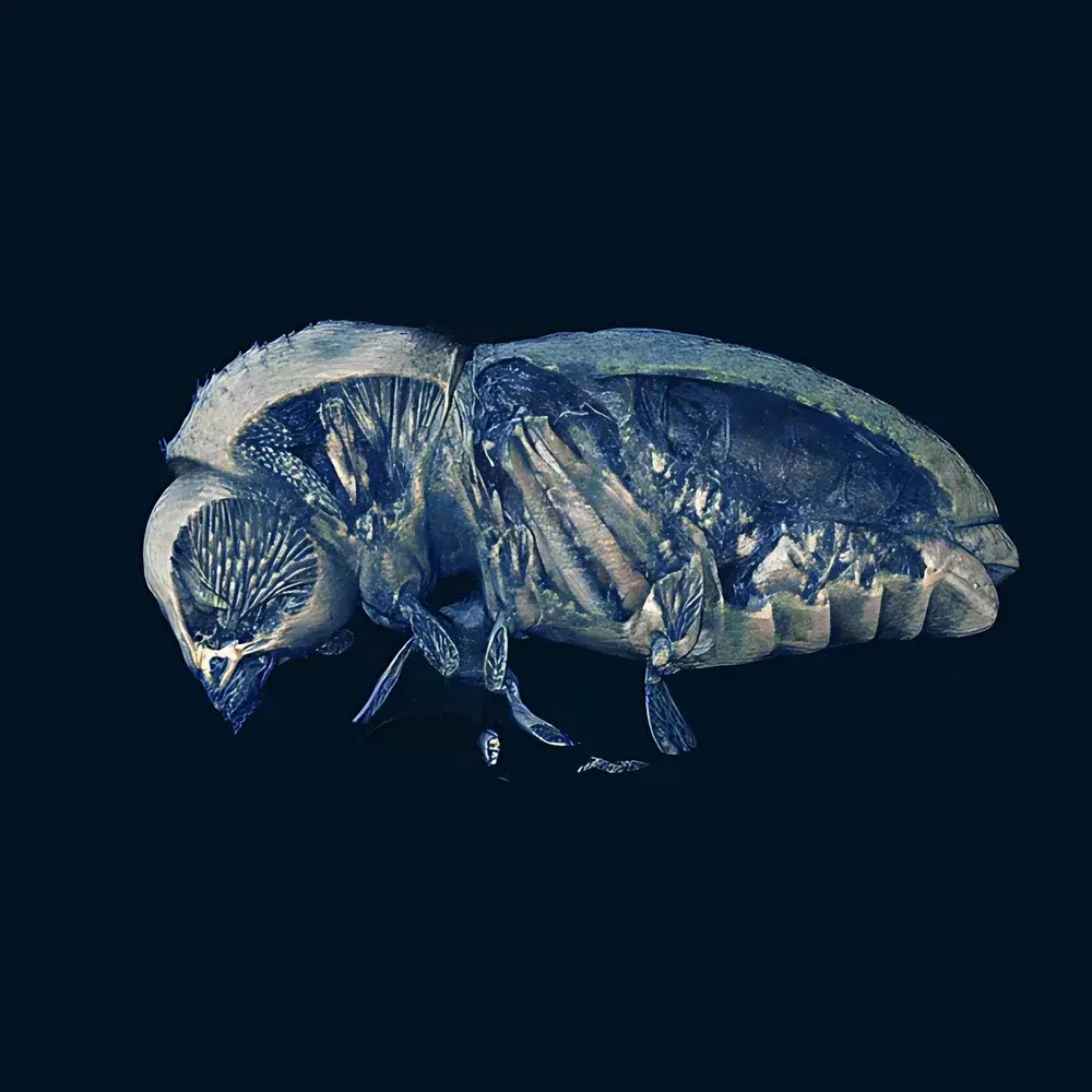
- Full workflow integration: from raw projection acquisition → 3D reconstruction → segmentation → morphometric quantification (porosity, thickness, connectivity, surface area/volume ratio) → STL mesh export for CAE simulation.
Sample Compatibility & Compliance
The N60 accommodates samples up to 20 kg and 60 mm in diameter × 36 mm height, supporting rigid, semi-rigid, and hydrated biological specimens when mounted in appropriate environmental enclosures (e.g., sealed vials or humidity-controlled stages). It complies with IEC 61000-6-3 (EMC emission standards) and meets EU Machinery Directive 2006/42/EC safety requirements. While not certified as a medical device, its imaging performance aligns with ASTM E1441-22 (Standard Guide for Computed Tomography) and ISO/IEC 17025:2017 principles for measurement traceability. Data provenance is maintained via embedded metadata (scan parameters, timestamps, operator ID), supporting GLP-compliant documentation workflows.
Software & Data Management
The Neoscan Acquisition & Analysis Suite is a unified Windows-based application offering intuitive tab-driven navigation across acquisition setup, live projection monitoring, reconstruction parameter tuning, and post-processing modules. All software licenses are perpetual and include free version upgrades for the life of the instrument. Reconstruction engines support both CPU- and CUDA-enabled GPU processing. Quantitative outputs adhere to DICOM-CT conventions where applicable and export natively to common formats: TIFF stacks, NIfTI, CSV (morphometric tables), and binary STL (watertight meshes). Audit trails record all user actions—including parameter changes and segmentation edits—in timestamped logs compliant with FDA 21 CFR Part 11 requirements for electronic records and signatures.
Applications
The N60 serves cross-disciplinary research and industrial QA/QC needs requiring sub-10 µm structural insight. In materials science, it enables pore network analysis in lithium-ion battery electrodes, defect mapping in additively manufactured Ti-6Al-4V components, and fiber orientation quantification in carbon-fiber-reinforced polymers. In life sciences, it supports murine skeletal phenotyping (trabecular thickness, bone volume fraction per BV/TV), dental implant-bone interface assessment, and plant root architecture modeling under controlled hydroponic conditions. Geoscientists apply it to quantify sandstone permeability predictors (pore throat distribution, coordination number), while pharmaceutical developers use it to correlate tablet compaction pressure with internal crack density and dissolution kinetics. Additional validated use cases include PCB solder void analysis, fossilized tissue preservation evaluation, and food matrix microstructure evolution during freeze-thaw cycling.
FAQ
What is the minimum achievable voxel size under optimal conditions?
The system achieves an effective isotropic voxel size of 8 µm when scanning objects near the center of the FOV at maximum magnification; actual resolution depends on source-detector distance, object size, and reconstruction kernel selection.
Does the N60 support time-lapse or in situ mechanical testing?
Yes—via optional motorized stage integration and external trigger synchronization (TTL input/output), enabling dynamic scans during uniaxial compression or thermal cycling experiments.
Can reconstructed volumes be exported for finite element analysis (FEA)?
Yes—binary STL meshes preserve topological integrity and are directly importable into ANSYS Mechanical, COMSOL Multiphysics, and Simulia Abaqus for stress-strain simulation.
Is remote operation supported?
The system supports secure remote desktop access and RESTful API integration for automated batch scanning protocols within enterprise LIMS or MES environments.
What maintenance is required for long-term calibration stability?
Annual geometric calibration using certified phantoms (e.g., NIST-traceable sphere arrays) and quarterly X-ray source output verification are recommended per ISO 12713:2020 guidelines for micro-CT systems.


