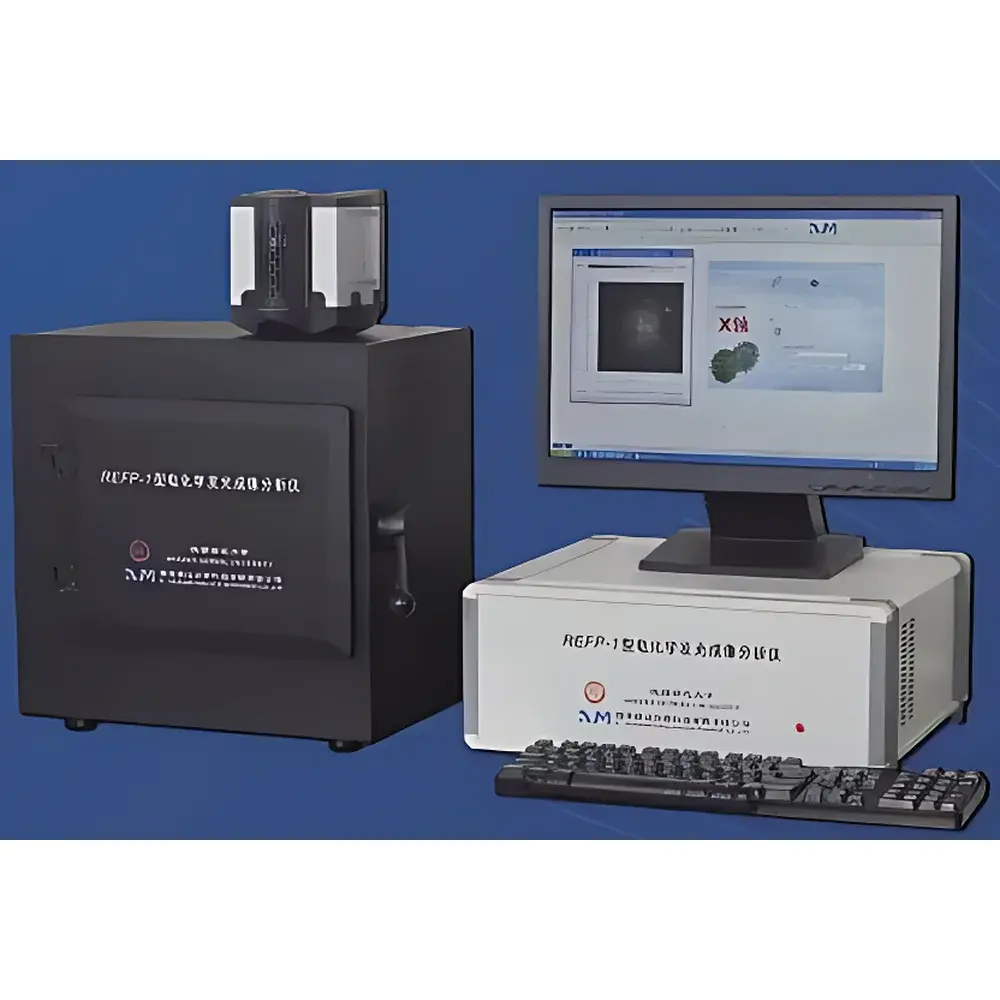
RIMAI REFP-1 Electrochemiluminescence Imaging Analyzer
| Origin | Shaanxi, China |
|---|---|
| Manufacturer Type | Authorized Distributor |
| Origin Category | Domestic (PRC) |
| Model | REFP-1 |
| Instrument Type | Optical Imaging System |
| Electrochemical Potential Range | −10 V to +10 V |
| Current Range | ±250 mA |
| Sensitivity | 1 × 10⁻⁹ A to 1 × 10⁻² A (8 decades) |
| Scan Rate | 0.0001–200 V/s |
| ECL Dynamic Range | >5 decades |
| ECL Wavelength Response | 230–920 nm |
| EMCCD Resolution | 512 × 512 pixels |
| Sensor Active Area | 8.192 mm × 8.192 mm |
| Readout Speed | ≤417 fps |
| Dark Current | 0.01 e⁻/pix/s |
| Detectable ECL Area | 5 mm × 5 mm |
| Optical Magnification Options | 4×, 10×, 20×, 40× |
| Integration Time | 1 ms–10 s |
| Sampling Rate | 1–200 Hz |
| Amplifier Drift | <0.05% |
| Signal Noise | 0.5 mV (peak-to-peak, gain = 1) |
Overview
The RIMAI REFP-1 Electrochemiluminescence Imaging Analyzer is a dedicated optical imaging platform engineered for quantitative, spatially resolved electrochemiluminescence (ECL) detection in life science and forensic research laboratories. Unlike conventional fluorescence or chemiluminescence imaging systems, the REFP-1 operates on the principle of electrode-confined ECL—where luminophores (e.g., Ru(bpy)₃²⁺ or luminol derivatives) undergo redox-triggered light emission directly at the electrode–solution interface. This eliminates photon diffusion artifacts, photobleaching, and external excitation source requirements, enabling intrinsic signal localization, sub-second temporal resolution, and exceptional signal-to-noise ratios. The system integrates dual-detection architecture: a high-gain electron-multiplying charge-coupled device (EMCCD) for wide-field, pixel-resolved ECL imaging, and a replaceable photomultiplier tube (PMT) module for ultra-low-light, single-point intensity quantification. Its design supports both static and dynamic ECL assays—including multi-electrode array mapping, spatially resolved biosensor arrays, and real-time kinetic profiling—under ambient or controlled environmental conditions.
Key Features
- Dual-detection modality: Interchangeable EMCCD imaging and PMT detection heads for flexible trade-offs between spatial resolution and ultimate sensitivity
- Integrated potentiostat with full electrochemical control: ±10 V potential range, ±250 mA current compliance, and programmable scan rates from 0.0001 to 200 V/s
- High-performance EMCCD sensor: 512 × 512 pixel resolution, <0.01 e⁻/pix/s dark current, and up to 417 frames per second readout—optimized for low-light ECL kinetics
- Precision XYZ motorized stage: Enables automated raster scanning across multi-electrode substrates and reproducible positioning of samples within the 5 mm × 5 mm active imaging field
- Multi-wavelength compatibility: Broad spectral response (230–920 nm) accommodates common ECL labels including ruthenium complexes, quantum dots, and acridinium esters
- Modular optical path: Interchangeable objectives (4×, 10×, 20×, 40×) allow scalable magnification without recalibration; no external excitation optics required
- Real-time data acquisition: 1–200 Hz sampling rate with auto-zeroing, drift-compensated amplification, and 5-decade linear dynamic range
Sample Compatibility & Compliance
The REFP-1 supports diverse sample formats including microelectrode arrays, printed circuit electrodes, tissue sections mounted on conductive ITO/glass slides, microfluidic ECL chips, and intact ex vivo organ preparations. It is compatible with aqueous and mixed-aqueous electrolytes (e.g., PBS, Tris-HCl, carbonate buffers), as well as organic co-solvents used in non-aqueous ECL systems. All electrochemical protocols adhere to ASTM E2916 (Standard Guide for Electrochemical Measurements in Corrosion Studies) and ISO 13843 (Electrochemical Sensors—Performance Requirements). Data acquisition and storage comply with GLP documentation standards, supporting audit trails, user authentication, and electronic signatures. While not pre-certified for clinical diagnostics under FDA 21 CFR Part 11, the software architecture permits configuration to meet traceability and data integrity requirements for regulated environments upon site-specific validation.
Software & Data Management
The REFP-1 is operated via RIMAI ECL-Studio™, a Windows-based application providing synchronized control of electrochemical waveform generation, stage movement, image capture, and PMT integration. The software enables batch acquisition of time-lapse ECL movies, region-of-interest (ROI) intensity profiling, false-color thermal mapping, and pixel-wise kinetic fitting (e.g., ECL rise/decay time constants). Raw image data are saved in TIFF format with embedded metadata (timestamp, electrode potential, integration time, objective ID); electrochemical traces export to ASCII or HDF5. Built-in calibration tools support intensity normalization using NIST-traceable luminance standards. Data processing modules include background subtraction, flat-field correction, and cross-channel registration for multi-label ECL assays. Export options include CSV, PNG, and PDF for direct integration into publication workflows.
Applications
- Clinical diagnostics: Multiplexed immunoassays for cardiac biomarkers, infectious disease antigens (e.g., SARS-CoV-2 nucleocapsid), and cancer-associated exosomal miRNAs
- Forensic science: Latent fingerprint visualization and chemical composition mapping via ECL-active metal ion chelation or antibody-conjugated probes
- Drug development: High-throughput screening of electroactive metabolites and doping agents (e.g., clenbuterol, ractopamine)
- Cellular analysis: Single-cell ECL imaging of membrane receptor dynamics, apoptosis-associated caspase activity, and intercellular signaling gradients
- Tissue-level imaging: Ex vivo ECL mapping of enzyme activity (e.g., alkaline phosphatase, glucose oxidase) in frozen sections and perfused organ slices
- Security screening: Detection of nitroaromatic explosives (e.g., TNT, RDX) and peroxide-based precursors using catalytic ECL amplification
- Basic research: Quantitative study of protein–protein interactions via proximity-induced ECL resonance energy transfer (PERET)
FAQ
What types of electrodes are compatible with the REFP-1?
Standard configurations support screen-printed carbon electrodes, gold microdisk arrays, indium tin oxide (ITO) glass slides, and custom planar electrode substrates up to 25 mm × 25 mm. Three-electrode cell setups (working, counter, reference) are fully supported.
Can the REFP-1 perform simultaneous multi-electrode ECL imaging?
Yes—the integrated potentiostat supports independent potential control of up to four working electrodes, and the EMCCD captures spatially resolved ECL from all active sites in parallel during a single exposure.
Is the EMCCD sensor cooled, and what is its quantum efficiency?
The EMCCD operates at −70 °C (thermoelectrically stabilized) and achieves >90% peak quantum efficiency in the 500–700 nm range, optimized for common Ru-based ECL emitters.
Does the system support kinetic ECL measurements with millisecond resolution?
Yes—using the PMT mode with 1 ms integration time and 1 kHz sampling, transient ECL responses (e.g., pulse-induced luminescence decay) can be resolved with sub-millisecond temporal fidelity.
What validation documentation is provided for regulatory use?
A comprehensive IQ/OQ protocol package is included, covering mechanical verification, electrochemical accuracy testing (per ASTM E2916), ECL intensity linearity (NIST SRM 2241 reference), and software functionality checks. Full 21 CFR Part 11 readiness requires customer-side risk assessment and procedural SOPs.


