
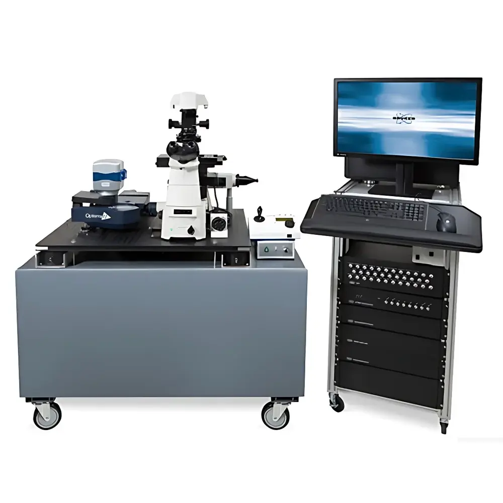
Bruker Opterra II Field-Scanning Confocal Imaging System
| Brand | Bruker |
|---|---|
| Origin | USA |
| Manufacturer Type | Authorized Distributor |
| Origin Category | Imported |
| Model | Opterra II |
| Price Range | USD 420,000 – 700,000 |
| Instrument Type | Optical Imaging System |
| Maximum Scan Resolution | 2048 × 2048 pixels |
| Frame Rate | up to 100 fps |
| Field of View | Objectives-dependent (e.g., 0.25 mm² at 60×, 1.0 mm² at 10×) |
| Sample Capacity | Single specimen stage with motorized XYZ + focus control |
| Detection Technology | EMCCD and sCMOS dual-path detection |
| Scanning Method | Parallel field-scanning via programmable slit/multi-spot illumination |
Overview
The Bruker Opterra II Field-Scanning Confocal Imaging System is a high-performance optical imaging platform engineered for quantitative, dynamic, and low-phototoxicity fluorescence microscopy in live biological specimens. Unlike conventional point-scanning confocal systems—whose serial acquisition limits temporal resolution and increases cumulative photon dose—the Opterra II implements a field-scanning architecture based on programmable multi-spot or slit illumination. This approach enables parallel optical sectioning across the entire field of view while preserving true confocality across a broad range of objective magnifications (10× to 100× oil). The system leverages dual high-sensitivity detection pathways: an electron-multiplying charge-coupled device (EMCCD) for ultra-low-light single-molecule applications and a scientific complementary metal-oxide-semiconductor (sCMOS) sensor for high-speed volumetric imaging. Its optical design complies with Köhler illumination principles and integrates aberration-corrected scan optics aligned to ISO 10110 standards for wavefront fidelity.
Key Features
- Field-scanning illumination engine supporting both multi-point and variable-width slit modes—enabling tunable optical sectioning thickness (0.3–5 µm) without mechanical pinhole replacement
- Simultaneous dual-channel detection with independently optimized gain, exposure, and readout timing for EMCCD and sCMOS sensors
- Motorized, encoded objective turret accommodating up to six objectives (including water-dipping and long-working-distance variants) with automatic magnification calibration
- Integrated piezo-driven Z-stage with sub-10 nm step resolution and closed-loop feedback for precise axial scanning and drift compensation
- Real-time hardware-based background subtraction and pixel-shift correction to mitigate thermal noise and detector non-uniformity
- Modular laser combiner supporting up to six solid-state lasers (405–785 nm), each with independent AOTF control and <1% power stability over 8 hours
Sample Compatibility & Compliance
The Opterra II accommodates standard glass-bottom dishes (35 mm, 50 mm), chambered coverslips, and custom-designed in vivo imaging chambers for rodent cranial window, spinal cord, or tumor xenograft preparations. It supports inverted and upright configurations via optional optical path modules. For regulatory environments, the system’s firmware includes audit-trail logging compliant with FDA 21 CFR Part 11 requirements, and acquisition metadata conforms to the OME-TIFF standard (Open Microscopy Environment) for interoperability with ImageJ/Fiji, Imaris, and commercial LIMS platforms. All optical components meet ISO 19012-1 (microscope nomenclature) and ISO 10934-1 (confocal performance verification) specifications. Routine validation protocols align with ASTM E2820-21 for confocal lateral resolution assessment and USP for fluorescence intensity linearity.
Software & Data Management
Acquisition and analysis are unified under Bruker’s proprietary Acquifer software suite, version 5.3+, which provides scriptable workflow automation (Python API), GPU-accelerated deconvolution (Richardson-Lucy with total variation regularization), and real-time rendering of 4D time-lapse stacks (x, y, z, t). Data export supports HCS (High-Content Screening) format for compatibility with Columbus and Harmony platforms. Raw image data are stored in vendor-neutral HDF5 containers with embedded CMTK (Computational Morphometry Toolkit) metadata tags. Software licensing includes annual updates, remote diagnostics, and GLP-compliant electronic lab notebook (ELN) integration via RESTful API endpoints.
Applications
The Opterra II serves as a core imaging modality in academic core facilities and pharmaceutical preclinical labs for longitudinal intravital imaging of immune cell trafficking, calcium dynamics in neuronal ensembles, mitochondrial fission/fusion kinetics, and angiogenic sprouting in transgenic zebrafish or murine models. Its speed-resolution balance makes it particularly suited for high-content screening of organoid drug responses, where >100 z-stacks per minute must be acquired across 96-well plates without photobleaching-induced artifacts. In developmental biology, the system enables synchronized multi-color time-lapse of gastrulation-stage embryos imaged through light-sheet-compatible clearing media (e.g., CUBIC or iDISCO+), with axial registration accuracy better than ±0.15 µm over 12-hour acquisitions.
FAQ
Does the Opterra II support spectral unmixing?
Yes—via sequential acquisition with tunable bandpass filters and reference spectra libraries for linear unmixing; full spectral detection requires optional prism-based spectrograph module.
Can the system be integrated into existing environmental control enclosures?
Yes—the main optical head has a footprint of 420 × 380 mm and interfaces with standard CO₂/O₂/temperature controllers via TTL and analog I/O ports.
Is training included with purchase?
Bruker provides onsite installation qualification (IQ), operational qualification (OQ), and two days of hands-on operator training covering acquisition optimization, maintenance routines, and basic quantitative analysis pipelines.
What service contracts are available?
Annual Platinum Support includes 24/7 remote diagnostics, priority on-site engineer dispatch (<48 hr response), preventive maintenance visits, and unlimited software upgrades.
How does field-scanning compare to spinning-disk confocal in thick-tissue imaging?
Unlike fixed-pinhole spinning disks, the Opterra II’s programmable slit geometry maintains consistent optical sectioning across varying refractive indices and depth—reducing out-of-focus flare by >40% in 100-µm-thick brain slices stained with Alexa Fluor 647.
Related Products


