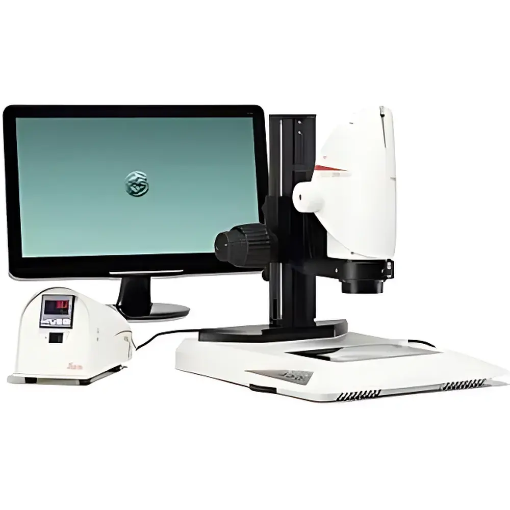
Leica DMS1000B Digital Stereo Microscope System
| Brand | Leica |
|---|---|
| Origin | Germany |
| Model | DMS1000B |
| Transmission Illumination Base Options | TL3000 ST and TL5000 Ergo |
| Max Optical Magnification | 300× |
| Camera Sensor | 5 MP CMOS |
| Video Output | Full HD (1920 × 1080) at 30 fps |
| Standalone Operation | Yes, with integrated SD card storage |
| Eyepiece-Free Imaging | Yes |
| Zoom Encoding | Yes, auto-calibrated scale overlay |
| Ergonomic Design | Optimized for laminar flow cabinet integration and multi-user workflows |
Overview
The Leica DMS1000B is a fully integrated, pre-configured digital stereo microscope system engineered for precision life science research environments—particularly those requiring strict contamination control, such as in vitro fertilization (IVF) laboratories, stem cell culture facilities, and embryology suites. Unlike traditional binocular stereo microscopes, the DMS1000B eliminates optical eyepieces entirely, relying instead on a high-fidelity digital imaging pathway: a calibrated zoom optics train coupled directly to an embedded 5-megapixel CMOS sensor. Its measurement principle is based on optically corrected stereoscopic imaging with real-time digital acquisition, enabling quantitative visual assessment without subjective observer variability. The system operates independently of a host PC—processing, previewing, capturing, and storing images and video directly onto a removable SD card—making it ideal for use inside ISO Class 5 laminar flow cabinets where cable management, space constraints, and biosafety protocols limit peripheral connectivity.
Key Features
- Standalone digital architecture: No external computer required for operation, image capture, or playback—reducing footprint and eliminating software compatibility dependencies.
- Integrated 5 MP CMOS camera delivering full HD (1920 × 1080) live video at 30 fps with low-latency rendering—critical for time-sensitive manipulations such as oocyte handling or blastocyst biopsy.
- Encoded zoom mechanism providing automatic magnification calibration and on-screen scale bar annotation for traceable, repeatable documentation compliant with GLP and ISO 13485-aligned workflows.
- Dual transmission illumination base compatibility: TL3000 ST (standard transillumination) and TL5000 Ergo (adjustable height, tilting stage with ergonomic controls), both optimized for high-contrast visualization of transparent or semi-opaque biological specimens.
- Infrared remote control interface supporting one-touch functions—including image/video capture, brightness adjustment, white balance, and display mode switching—enabling glove-compatible operation within containment enclosures.
- Ergonomically optimized display positioning: The 19″–24″ external monitor (sold separately) mounts flexibly to accommodate seated or standing users, eliminating need for individual eyepiece refocusing—a key advantage in shared-lab or clinical rotation settings.
Sample Compatibility & Compliance
The DMS1000B supports non-invasive, high-resolution observation of live and fixed biological samples including zygotes, embryos, oocytes, tissue explants, and microinjected constructs. Its sealed optical path and absence of oculars prevent aerosol-mediated cross-contamination, aligning with WHO Good Manufacturing Practice (GMP) recommendations for assisted reproductive technology (ART) labs. The system meets CE marking requirements under the EU Medical Device Regulation (MDR 2017/745) when used as an ancillary device in diagnostic or therapeutic procedures. While not a Class IIa medical device itself, its design facilitates compliance with ISO 15189 (medical laboratories) and ISO 13485 (quality management for medical devices) through audit-ready metadata logging (timestamped captures with embedded magnification and illumination settings).
Software & Data Management
All image and video files are saved in standard DICOM-compliant JPEG or MP4 formats directly to SDHC cards (up to 128 GB), ensuring interoperability with hospital PACS systems and LIMS platforms. Optional Leica Application Suite (LAS X) Core software enables advanced post-acquisition analysis—including region-of-interest (ROI) measurement, contrast enhancement, multi-frame averaging, and export to CSV for statistical processing. Audit trail functionality records operator ID (via optional RFID login), capture timestamp, magnification, illumination intensity, and focus position—supporting FDA 21 CFR Part 11 requirements when deployed in regulated GxP environments.
Applications
- In vitro fertilization (IVF) and intracytoplasmic sperm injection (ICSI) procedures, including embryo grading per Istanbul consensus criteria.
- Stem cell colony monitoring and passaging under sterile laminar airflow.
- Microdissection of neural tissues, organoids, or early-stage embryos.
- Quality control of microfluidic devices and lab-on-chip substrates.
- Training and tele-mentoring in reproductive medicine, where real-time screen sharing replaces direct ocular supervision.
FAQ
Does the DMS1000B require a computer to operate?
No—the system runs autonomously with built-in firmware for live imaging, capture, playback, and SD card storage.
Can I use my existing Leica TL base with the DMS1000B?
Yes, the DMS1000B is mechanically and electronically compatible with both the TL3000 ST and TL5000 Ergo transmission bases.
Is the magnification scale bar calibrated per zoom setting?
Yes—encoded zoom ensures pixel-to-micron calibration is automatically applied and displayed in real time.
What file formats are supported for export?
JPEG (still images), MP4 (H.264-encoded video), and BMP; all embed EXIF metadata including magnification, exposure, and timestamp.
How is user authentication handled in regulated environments?
Optional RFID reader integration enables operator identification, with session logs stored alongside captured media for 21 CFR Part 11 compliance.


