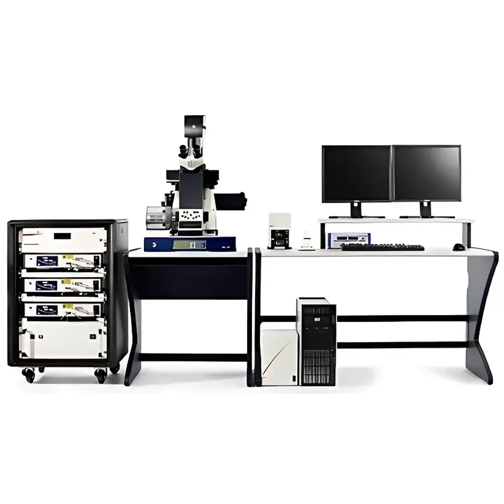
Leica SR GSD Super-Resolution Ground State Depletion Microscope
| Brand | Leica |
|---|---|
| Origin | Germany |
| Model | Leica SR GSD |
| Laser Wavelengths | 405 nm, 488 nm, 532 nm, 642 nm |
| Laser Power | 300–1000 mW per line |
| Lateral Drift Stability | <20 nm / 10 min (SuMo Piezo Stage) |
| Resolution (GSDIM mode) | ≤20 nm (lateral) |
| Compatible Fluorophores | Alexa Fluor® 488/532/546/647, Rhodamine-6G, Atto 488/532/565/568, YFP, EYFP |
| Imaging Modes | Widefield, TIRF, High-Speed Multichannel Fluorescence, Temperature-Controlled Live-Cell Imaging |
| Base Platform | Leica DMI6000 B Inverted Microscope with AM TIRF MC System |
Overview
The Leica SR GSD Super-Resolution Ground State Depletion Microscope is a purpose-engineered optical platform for single-molecule localization microscopy (SMLM) based on the GSDIM (Ground State Depletion followed by Individual Molecule return) principle. Unlike diffraction-limited widefield or confocal systems, the Leica SR GSD exploits reversible photoswitching of standard organic fluorophores into a long-lived dark state—typically the triplet or radical ion state—enabling stochastic activation and precise centroid localization of individual emitters. This physical mechanism, originally developed at the Max Planck Institute for Biophysical Chemistry (Göttingen) and licensed exclusively to Leica Microsystems, delivers lateral resolution down to ≤20 nm under physiological conditions. The system integrates seamlessly with the Leica DMI6000 B inverted microscope and the AM TIRF MC module, providing a mechanically stable, modular architecture optimized for both fixed-sample nanoscopy and dynamic live-cell imaging. Its design prioritizes experimental reproducibility, minimal workflow disruption, and compatibility with routine immunolabeling protocols—eliminating the need for specialized dyes, buffer formulations, or post-acquisition computational corrections not grounded in first-principles photophysics.
Key Features
- GSDIM-optimized optical path: Dedicated high-numerical-aperture objective coupling, optimized dichroic filters, and quad-band emission detection ensure maximal photon collection efficiency and minimal crosstalk across 405/488/532/642 nm excitation channels.
- SuMo piezo-driven stage: A pressure-driven, closed-loop motorized stage delivering sub-20 nm lateral drift stability over 10-minute acquisition windows—critical for accurate molecular coordinate reconstruction in dense labeling environments.
- Real-time localization rendering: On-the-fly image reconstruction engine enables immediate visualization of accumulating localizations during acquisition, allowing adaptive termination or extension of frame counts without post-hoc processing latency.
- Multimodal imaging integration: Native support for simultaneous TIRF, epifluorescence, and temperature-controlled (20–40 °C) live-cell imaging within the same optical train—ensuring consistent alignment and calibration across modalities.
- Standard fluorophore compatibility: Validated performance with widely adopted probes including Alexa Fluor® 488, 532, 546, 647; Atto dyes (488, 532, 565, 568); Rhodamine-6G; and fluorescent proteins (YFP, EYFP), preserving established sample preparation workflows.
Sample Compatibility & Compliance
The Leica SR GSD accommodates standard glass-bottom dishes (e.g., MatTek, Ibidi), coverslips (No. 1.5H), and custom chambered substrates compatible with high-NA oil or water immersion objectives (63×–160×). It supports both chemically fixed specimens and live cells expressing genetically encoded tags or labeled via antibody-based protocols. All hardware and firmware components comply with IEC 61000-6-3 (EMC emission) and IEC 61000-6-2 (immunity) standards. Data acquisition logs—including timestamped laser power, stage position, exposure duration, and filter wheel state—are recorded in vendor-neutral HDF5 format with embedded metadata, supporting traceability requirements under GLP and ISO/IEC 17025 frameworks. While not FDA 21 CFR Part 11-certified out-of-the-box, audit trail functionality (user login, parameter change history, raw data integrity checksums) can be enabled via optional Leica Application Suite X (LAS X) modules for regulated environments.
Software & Data Management
Control and analysis are performed using Leica Application Suite X (LAS X) software, which provides a unified interface for instrument orchestration, real-time GSDIM reconstruction, and quantitative colocalization analysis. The software implements maximum-likelihood estimation (MLE) and Gaussian fitting algorithms compliant with the International Standard ISO/IEC 17025:2017 Annex A.2 for measurement uncertainty propagation in localization data. Raw camera frames, intermediate localization lists (.csv/.xml), and rendered super-resolution images (.tif/.ome.tiff) are stored with FAIR-compliant metadata (OME-XML schema). LAS X supports batch processing pipelines exportable as Python scripts, enabling integration with open-source tools such as ThunderSTORM or Picasso for secondary validation. Data export adheres to MIAP (Minimum Information About a Super-Resolution Microscopy Experiment) guidelines, facilitating publication-ready figure generation and repository submission (e.g., BioImage Archive).
Applications
- Nuclear pore complex (NPC) ultrastructure mapping in Ptk2 cells using anti-NUP153/Alexa Fluor® 532 labeling.
- Microtubule lattice organization in MDCK and RCC-FG1 cells resolved at sub-20 nm precision using dual-color Alexa Fluor® 488/647 labeling.
- Golgi apparatus nanoscale architecture in B16 melanoma cells via EYFP-tagged β-1,4-galactosyltransferase.
- Dynamic clathrin-coated pit assembly kinetics under TIRF-GSDIM dual-mode acquisition.
- Quantitative co-distribution analysis of synaptic proteins in primary neuronal cultures with <15 nm positional confidence intervals.
FAQ
What is the fundamental physical principle underlying GSDIM imaging on the Leica SR GSD?
GSDIM relies on transient depletion of fluorophores into a long-lived non-fluorescent ground-state sublevel (e.g., triplet or radical anion state) using high-intensity 405 nm light, followed by stochastic return to the emissive singlet state. This enables temporally isolated emission events suitable for single-molecule centroid localization.
Can the Leica SR GSD perform live-cell super-resolution imaging?
Yes—when combined with temperature control (20–40 °C), low-phototoxicity illumination schemes, and fast sCMOS cameras, the system supports time-lapse GSDIM of cellular structures with frame rates up to 10 Hz and localization precision <30 nm over extended durations.
Is special sample preparation required compared to conventional fluorescence microscopy?
No—standard immunostaining protocols using common fixatives (PFA, methanol), blocking agents (BSA, serum), and commercial antibodies conjugated to validated fluorophores are fully compatible.
How does the SuMo stage achieve sub-20 nm drift stability?
It employs a proprietary pressure-driven piezoelectric actuator with integrated capacitive position sensing and real-time feedback correction, decoupling mechanical drift from thermal expansion and acoustic perturbations.
What file formats are generated during GSDIM acquisition and analysis?
Raw EMCCD/sCMOS frames (.tif), localization tables (.csv/.xml), reconstructed super-resolution images (.ome.tiff), and comprehensive metadata (OME-XML) are natively supported and exportable for third-party analysis.


