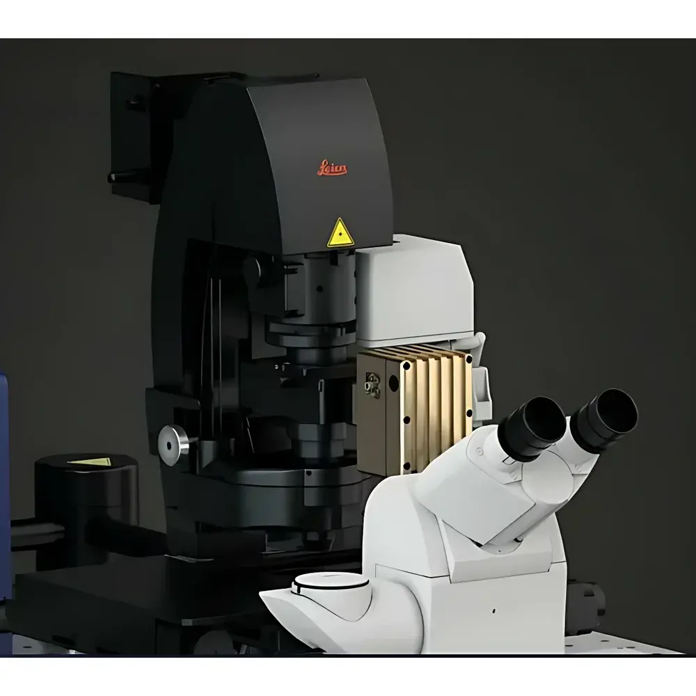
Leica STELLARIS CRS Coherent Raman Scattering Microscope
| Brand | Leica |
|---|---|
| Origin | Germany |
| Model | STELLARIS CRS |
| Instrument Type | Point-Scanning Confocal Microscope |
| Excitation | Near-Infrared (NIR) Lasers |
| Imaging Modalities | Stimulated Raman Scattering (SRS), Coherent Anti-Stokes Raman Scattering (CARS), Multiphoton Fluorescence, Second Harmonic Generation (SHG), FLIM |
| Spatial Resolution | Subcellular (≤ 300 nm lateral, ≤ 1 µm axial) |
| Detection | Spectral Unmixing, TauSense Lifetime-Gated Acquisition |
| Software Platform | LAS X with ImageCompass Interface |
| Compliance | Designed for GLP/GMP-aligned workflows |
Overview
The Leica STELLARIS CRS Coherent Raman Scattering Microscope is a fully integrated, point-scanning confocal platform engineered for label-free, chemically specific imaging at subcellular resolution. Unlike conventional fluorescence microscopy—which relies on exogenous dyes or genetically encoded fluorophores—the STELLARIS CRS leverages intrinsic molecular vibrations to generate contrast. It operates on the physical principles of coherent Raman scattering, primarily Stimulated Raman Scattering (SRS) and Coherent Anti-Stokes Raman Scattering (CARS), both nonlinear optical processes that occur only within the diffraction-limited focal volume. This enables true optical sectioning without post-acquisition deconvolution, delivering native 3D chemical contrast in thick, unprocessed biological specimens—including live tissues, organoids, and small model organisms. The system utilizes near-infrared (NIR) excitation lasers, minimizing photon absorption, scattering, and phototoxicity—critical for long-term live-cell and developmental dynamics studies under near-physiological conditions.
Key Features
- Label-free chemical imaging based on intrinsic vibrational signatures of biomolecules (e.g., CH2, CH3, C=O, C=C bonds), eliminating artifacts from staining, fixation, or photobleaching
- Simultaneous multimodal acquisition combining SRS, CARS, multiphoton fluorescence, SHG, and fluorescence lifetime imaging (FLIM) within a single optical path
- TauSense technology for temporal discrimination between Raman signals and autofluorescence, enabling robust separation of CARS and endogenous fluorescence in heterogeneous samples
- Resonant scanning architecture supporting video-rate imaging (up to 30 fps at full frame) for dynamic metabolic monitoring and real-time subcellular trafficking
- Sub-micron axial resolution and <300 nm lateral resolution across visible to NIR spectral ranges, validated per ISO 19012-1 for confocal performance
- Integrated LAS X software with ImageCompass user interface—providing guided workflow setup, intuitive laser parameter control, and one-click switching between bond-specific, spectral, and hybrid imaging modes
Sample Compatibility & Compliance
The STELLARIS CRS is optimized for native-state imaging of delicate, light-sensitive biological systems. Its NIR excitation (typically 700–1100 nm) ensures deep penetration and minimal thermal load in aqueous environments, making it suitable for extended time-lapse observation of primary neurons, embryonic tissues, and perfused organ slices. No sample pre-treatment—such as fixation, dehydration, or embedding—is required. The system complies with design principles aligned with Good Laboratory Practice (GLP) and Good Manufacturing Practice (GMP) frameworks: audit-trail logging, user access controls, and electronic signature support are configurable in LAS X to meet regulatory expectations under FDA 21 CFR Part 11. While not certified as a medical device, its architecture supports traceable, reproducible data generation essential for preclinical pharmacokinetic, toxicological, and mechanistic studies adhering to OECD, ISO/IEC 17025, and ASTM E2554 standards.
Software & Data Management
LAS X serves as the unified acquisition and analysis environment for the STELLARIS CRS. It natively supports quantitative spectral unmixing, ratiometric analysis (e.g., lipid/protein ratio mapping), and hyperspectral data cubes (wavenumber-resolved SRS stacks). All raw intensity, lifetime, and spectral metadata are stored in vendor-neutral HDF5 format with embedded calibration references. ImageCompass provides context-aware guidance—automatically suggesting optimal laser power, dwell time, and spectral window based on selected molecular target (e.g., 2845 cm−1 for CH2 symmetric stretch). Batch processing pipelines enable automated registration of multimodal datasets (e.g., coregistering SRS lipid maps with FLIM-based NAD(P)H decay profiles), facilitating correlative biochemical phenotyping. Data export conforms to MIAP (Minimum Information About a Photonics Experiment) guidelines, ensuring interoperability with FAIR-aligned repositories.
Applications
The STELLARIS CRS addresses fundamental challenges in translational life sciences where molecular identity—not just localization—is decisive. Key use cases include: real-time tracking of lipid droplet biogenesis and turnover in adipocytes; spatially resolved monitoring of drug distribution and metabolism in tumor spheroids; label-free assessment of myelin integrity in neurodegenerative models; quantification of collagen cross-linking heterogeneity in fibrotic tissue; and simultaneous visualization of structural (SHG), metabolic (SRS), and functional (FLIM) parameters in developing zebrafish embryos. Its ability to acquire chemically specific, volumetric, time-resolved data without perturbing homeostasis makes it particularly valuable for longitudinal studies of cellular aging, stem cell differentiation, and host–pathogen interactions—where dye-induced stress or photodamage would compromise physiological relevance.
FAQ
How does SRS differ from spontaneous Raman microscopy?
SRS employs two synchronized, ultrafast NIR lasers (pump and Stokes) to drive stimulated emission at a specific Raman shift, yielding orders-of-magnitude higher signal-to-noise than spontaneous Raman—and enabling video-rate imaging without signal averaging.
Can STELLARIS CRS replace fluorescence labeling entirely?
It complements—but does not universally replace—fluorescence. While SRS excels at imaging abundant endogenous molecules (lipids, proteins, water), low-concentration targets (e.g., specific receptors) still require fluorescent probes; however, multimodal integration allows co-registration of both contrast mechanisms.
Is spectral calibration traceable to international standards?
Yes. LAS X includes built-in polystyrene and cyclohexane reference spectra for wavenumber calibration, traceable to NIST SRM 2241 and ISO 8601-1 certified Raman shift standards.
What maintenance protocols ensure long-term measurement stability?
Leica recommends quarterly alignment verification using the internal reference channel and annual factory recalibration of laser wavelength, pulse duration, and detector quantum efficiency—documented per ISO/IEC 17025 calibration records.
Does the system support third-party analysis tools?
Raw HDF5 datasets are compatible with Python (via h5py), MATLAB, and open-source platforms including QuantiFly and SRSAnalyzer, enabling custom spectral fitting, machine learning segmentation, and cross-platform reproducibility validation.


