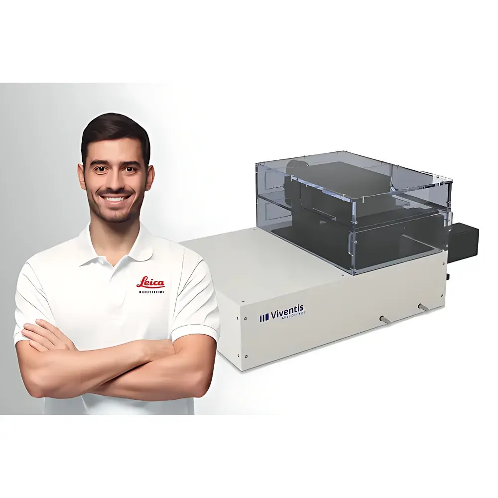
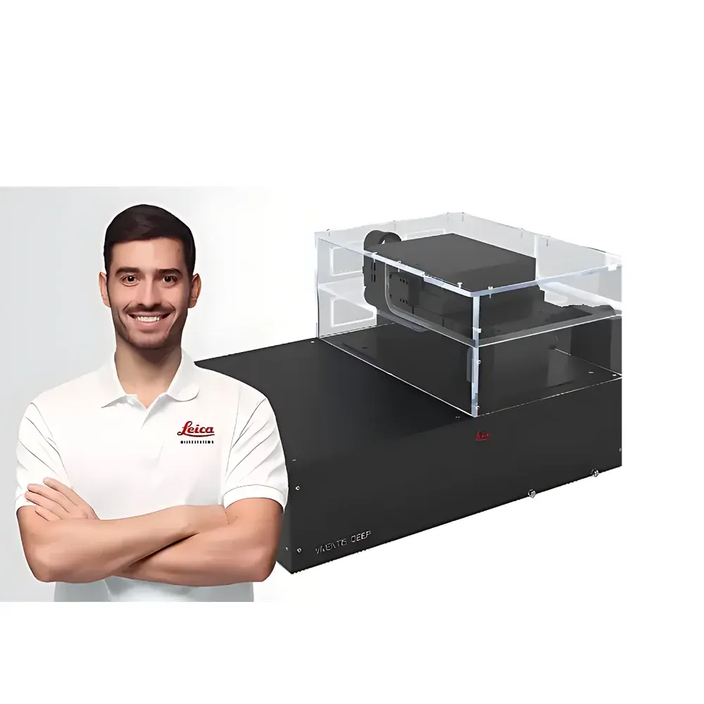
Leica Viventis Deep Dual-View Light Sheet Fluorescence Microscope
| Brand | Leica |
|---|---|
| Origin | Austria |
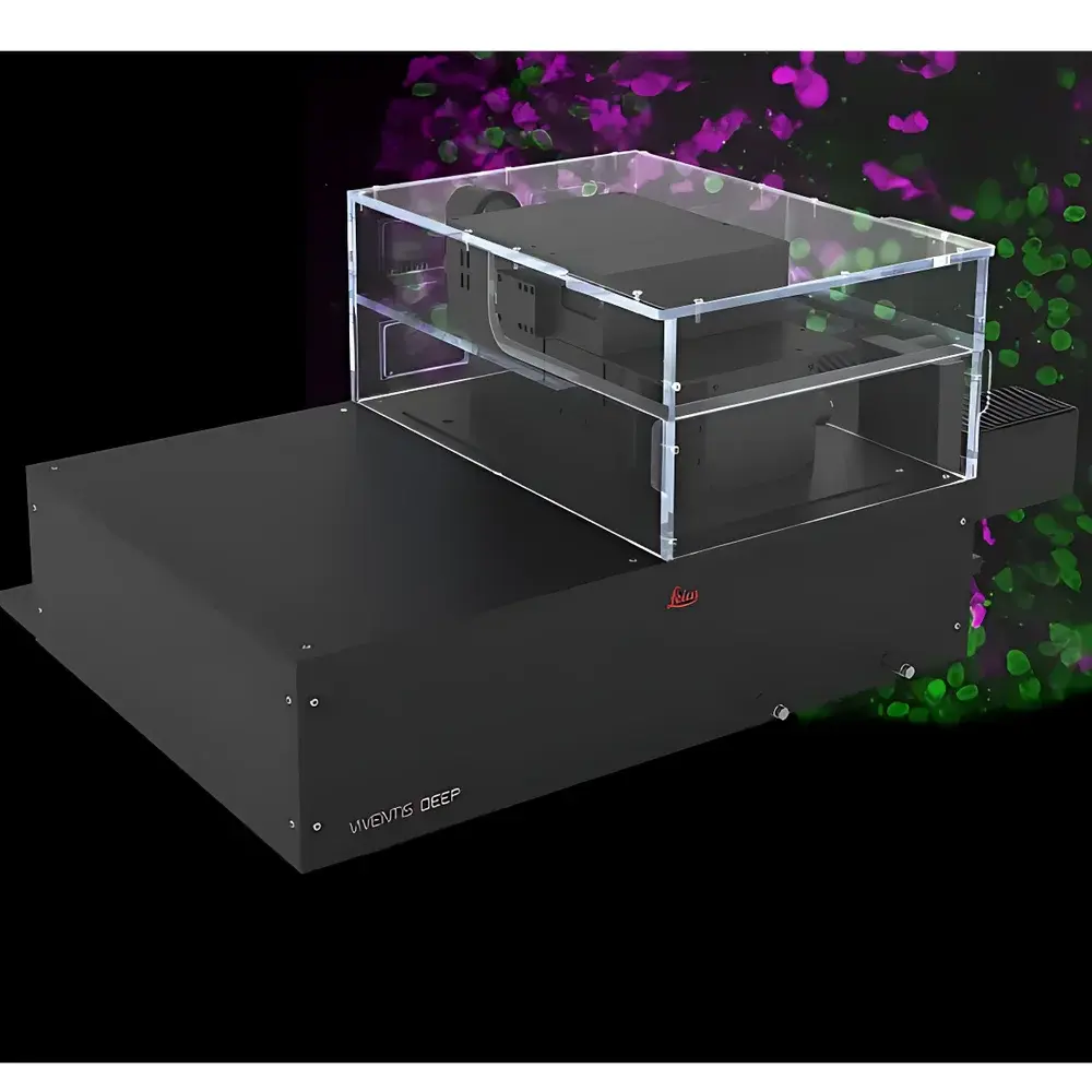
| Model | Viventis Deep |
| Light Sheet Technology | Dual-side illumination with scanned Gaussian light sheet |
| Imaging Resolution | 406 nm (with 16× objective), 260 nm (with 25× objective) |
| Field of View | 900 µm (16×), 596 µm (25×) |
| Maximum Sample Size | >50 mm |
| Laser Wavelengths | 405 nm, 488 nm, 561 nm, 638 nm |
| Objective Lenses | 16× and 25× high-NA water-dipping objectives |
| Illumination Mode | Dual-side illumination |
Overview
The Leica Viventis Deep Dual-View Light Sheet Fluorescence Microscope is an advanced selective-plane illumination platform engineered for high-fidelity, long-term volumetric imaging of large, optically challenging biological specimens. Operating on the principle of orthogonal light sheet generation—where a thin, scanned Gaussian light sheet illuminates only the focal plane of detection—the system minimizes out-of-focus excitation, phototoxicity, and photobleaching. Its dual-view architecture integrates two independent illumination-detection paths aligned at 90°, enabling simultaneous acquisition from complementary perspectives without mechanical repositioning. This design supports intrinsic optical sectioning, subcellular resolution across millimeter-scale volumes, and quantitative reconstruction of dynamic processes in live embryos, organoids, cleared tissues, and whole-mount preparations.
Key Features
- Dual-side scanned Gaussian light sheet illumination: Enables uniform, diffraction-limited optical sectioning while maintaining low photodose across extended time-lapse experiments.
- Two high-numerical-aperture water-dipping objectives (16× and 25×): Deliver lateral resolutions of 406 nm and 260 nm respectively, validated per ISO 19012-1 standards for point-spread function characterization.
- Top-access open-sample chamber: Accommodates specimens up to >50 mm in height, compatible with custom perfusion systems, environmental control units (temperature, CO₂, humidity), and motorized multi-position sample staging.
- Integrated Class 3B laser engine: Four solid-state lasers (405 nm, 488 nm, 561 nm, 638 nm) with optional wavelength expansion; all diodes are thermally stabilized and fiber-coupled for beam homogeneity and long-term power stability (±1.5% over 8 h).
- Synchronized dual-camera acquisition: Independent sCMOS detectors capture orthogonal views in real time, supporting computational fusion, self-consistent registration, and artifact suppression via joint deconvolution algorithms.
- Modular optical path design: Supports third-party add-ons including adaptive optics modules, spectral unmixing filters, and time-gated fluorescence lifetime extensions (FLIM-ready interface).
Sample Compatibility & Compliance
The Viventis Deep is validated for use with diverse specimen types—including zebrafish and Drosophila embryos, mouse brain organoids, cleared murine organs (e.g., CLARITY-, CUBIC-, or SHIELD-processed), and thick plant meristems—without requiring physical sectioning. Its open architecture complies with ISO/IEC 17025 requirements for calibration traceability and supports GLP/GMP-aligned workflows through audit-trail-enabled acquisition logging. All laser safety protocols conform to IEC 60825-1:2014 and ANSI Z136.1-2022 standards. The system meets electromagnetic compatibility (EMC) requirements per EN 61326-1:2013 and is CE-marked for research use only (RUO) in the EU and US markets.
Software & Data Management
Acquisition and reconstruction are managed via Leica Application Suite X (LAS X) Light Sheet Edition v4.1+, which provides automated multiview registration, GPU-accelerated deconvolution (Richardson-Lucy algorithm), and native support for HDF5-based data containers compliant with the OME-NGFF specification. The software enforces metadata embedding per MIAME and MINSEQE guidelines, includes built-in DICOM-SR export for cross-platform interoperability, and supports 21 CFR Part 11-compliant user authentication, electronic signatures, and immutable audit trails when deployed in regulated environments. Raw datasets are structured using the Zarr format for scalable cloud ingestion and integration with Python-based analysis pipelines (e.g., napari, ClearMap2, BigStitcher).
Applications
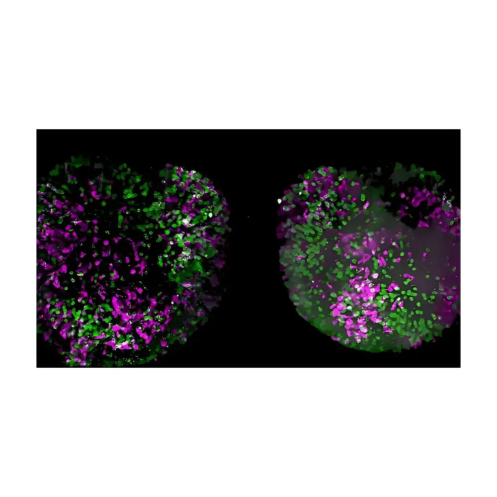
- Longitudinal developmental biology: Tracking cell lineage, morphogenetic movements, and tissue folding dynamics in intact vertebrate embryos over 48–72 h at ≤10 s temporal resolution.
- Neurovascular mapping: Volumetric imaging of fluorescently labeled vasculature and microglia in centimeter-scale cleared brains with isotropic voxel sampling down to 0.35 µm³.
- Organoid maturation studies: Quantifying lumen formation, polarization markers, and calcium signaling gradients across heterogeneous 3D cultures under physiological perfusion.
- Plant phenotyping: Time-resolved imaging of root gravitropism, vascular patterning, and pathogen response in whole seedlings up to 8 cm tall.
- Multimodal correlative workflows: Seamless integration with serial block-face SEM or MALDI-MSI datasets via common coordinate frameworks (e.g., BigWarp, TrakEM2).
FAQ
What sample mounting configurations are supported?
Standard configurations include agarose-embedded specimens in glass capillaries, custom 3D-printed holders for upright or inverted orientation, and magnetic levitation stages for contact-free positioning. Optional vacuum-sealed chambers enable long-term imaging under controlled gas atmospheres.
Is the system compatible with adaptive optics correction?
Yes—the optical train includes a dedicated pupil plane port for insertion of deformable mirrors or spatial light modulators, with software-integrated wavefront sensing via Shack-Hartmann sensor integration.
How is multiview data registered and fused?
LAS X implements intensity-based rigid and non-rigid registration using mutual information optimization, followed by weighted averaging or maximum-intensity projection with view-specific confidence masking.
Can the system be integrated into automated core facilities?
Fully supported via RESTful API endpoints for remote scheduling, acquisition triggering, and metadata push to LIMS platforms (e.g., LabVantage, STARLIMS); hardware-level I/O triggers available for synchronization with external stimulators or electrophysiology rigs.
What maintenance and service options are available globally?
Leica offers tiered service plans including Preventive Maintenance Contracts (PMCs), On-Site Calibration Services traceable to PTB/NIST standards, and 24/7 OneCall engineering support staffed by PhD-level application scientists—with regional spare parts hubs in Singapore, Frankfurt, Boston, São Paulo, and Dubai.
Related Products


