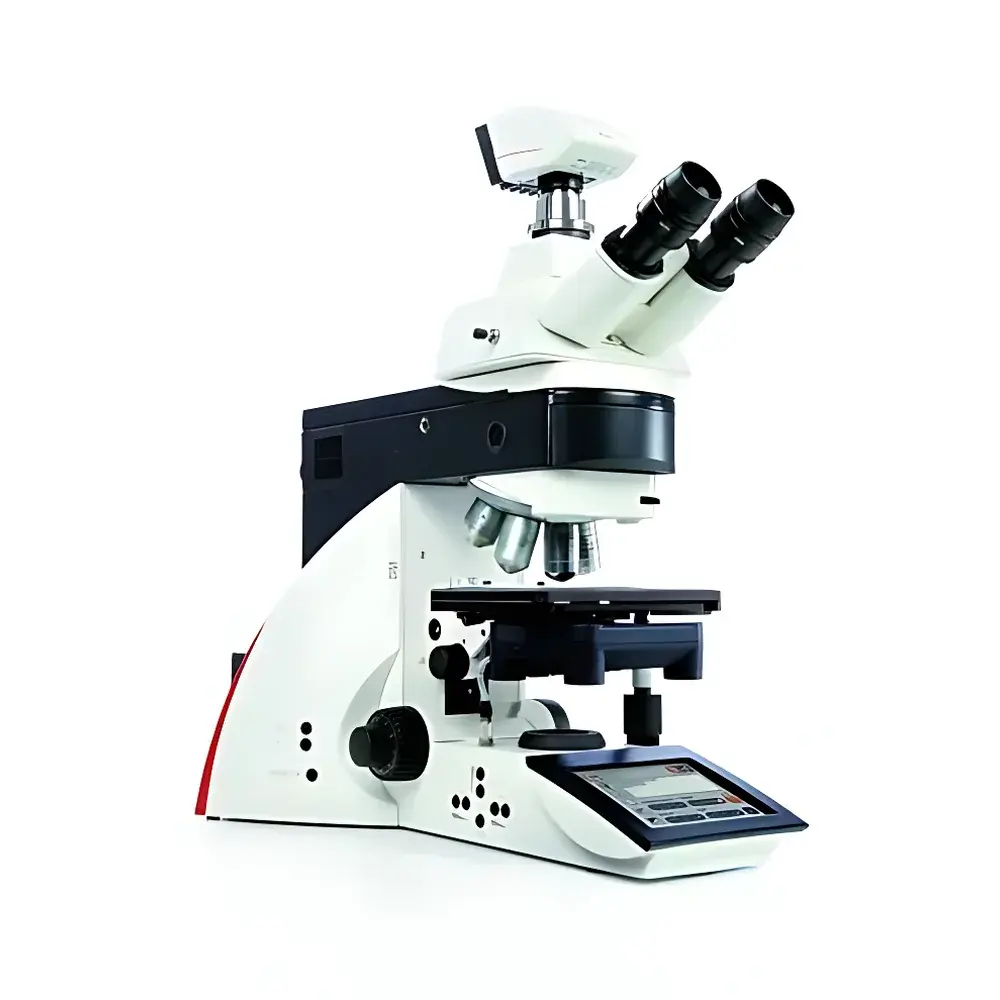
Leica DM5000 B Automated Upright Microscope System for Life Science Research
| Brand | Leica |
|---|---|
| Origin | Germany |
| Model | DM5000 B |
| Configuration | Motorized DIC, Touchscreen Control, 7-Position Encoded Nosepiece, Manual Z-Focus, 5- or 8-Position Motorized Fluorescence Filter Turret, Fluorescence Intensity Manager (FIM) |
| Compliance | Designed for GLP/GMP-aligned workflows, compatible with ISO 13485–informed lab environments |
| Software Integration | Leica Application Suite X (LAS X) ready |
Overview
The Leica DM5000 B is an automated upright microscope system engineered for rigorous life science research applications—particularly live-cell imaging, morphological phenotyping, and long-term time-lapse studies of adherent and suspension cultures. Built on Leica’s proven optical architecture, the DM5000 B employs Köhler illumination-coupled transmitted-light optics and high-NA plan-apochromat objectives to deliver diffraction-limited resolution across brightfield, darkfield, phase contrast, and polarized light modalities. Its core differentiating capability lies in fully encoded, motorized Differential Interference Contrast (DIC) optics—enabling rapid, repeatable switching between contrast modes without mechanical realignment. Unlike manual DIC systems requiring prism repositioning and bias adjustment, the DM5000 B integrates stepper-motor-driven Nomarski prisms and condenser alignment modules, ensuring precise, user-independent DIC contrast generation at any magnification. The system’s upright configuration supports standard Petri dishes, multi-well plates, and custom culture chambers—making it suitable for both fixed-tissue histology and dynamic in vitro assays under physiological conditions.
Key Features
- 7-position encoded nosepiece with automatic objective recognition and metadata tagging for traceable acquisition parameters
- Motorized, software-synchronized DIC module with integrated Wollaston prism positioning and bias control—eliminating manual fine-tuning
- Motorized fluorescence turret accommodating either 5 or 8 filter sets; each position auto-registered and recallable via LAS X
- Leica Fluorescence Intensity Manager (FIM): a hardware-software closed-loop system that dynamically modulates lamp intensity or LED output to maintain consistent photon flux across channels and sessions
- Full-color 7-inch capacitive touchscreen interface with context-sensitive controls, real-time parameter display, and intuitive workflow navigation
- Dedicated programmable shortcut keys adjacent to focus knobs—configurable per user profile or assay protocol (e.g., “DIC ON → 40x → Capture”)
- Manual Z-axis focusing mechanism with dual-speed coaxial focus drive and fine-focus vernier scale for tactile precision during manual observation
Sample Compatibility & Compliance
The DM5000 B accommodates standard microscopy sample formats including glass slides, coverslips (No. 1.5), 6–96-well tissue culture plates, and custom-stage inserts for perfusion chambers or environmental control enclosures. Its stage design supports optional motorized XY translation and Z-height calibration for multi-position time-series. From a regulatory standpoint, the system supports audit-trail-capable operation when paired with LAS X software configured under 21 CFR Part 11-compliant settings (electronic signatures, session logging, parameter change history). While not a medical device itself, its optical performance aligns with ISO 10934-1 (microscopy terminology) and ASTM E2101 (standard practice for fluorescence microscopy quantification), facilitating data acceptance in peer-reviewed publications and preclinical reporting frameworks.
Software & Data Management
The DM5000 B operates natively with Leica Application Suite X (LAS X), a modular platform supporting multi-channel fluorescence acquisition, Z-stack reconstruction, spectral unmixing, and quantitative intensity profiling. All hardware states—including objective ID, DIC prism position, fluorescence filter selection, exposure time, gain, and FIM-set intensity—are embedded into TIFF and LIF metadata headers. LAS X enables protocol-based acquisition: users define complete imaging sequences (e.g., “DIC @ 20x + GFP @ 40x + TRITC @ 63x”) and save them as executable methods. These methods are exportable, version-controlled, and reproducible across identical DM5000 B installations—ensuring inter-laboratory comparability. Raw image data can be exported in OME-TIFF format for integration with open-source analysis pipelines (e.g., Fiji/ImageJ, QuPath, or Python-based scikit-image workflows).
Applications
- Longitudinal monitoring of cell migration, mitosis, and organelle dynamics in primary neurons or stem-cell-derived organoids
- Quantitative assessment of cytoskeletal reorganization using phalloidin-AlexaFluor staining under DIC/fluorescence overlay
- High-fidelity documentation of tissue section morphology in developmental biology and toxicology screening
- Multi-parametric phenotyping in CRISPR-edited cell lines—correlating morphology (DIC), nuclear localization (Hoechst), and reporter expression (GFP/RFP)
- Standardized quality control of bioprocess-relevant cell cultures (e.g., viability, confluence, morphology) prior to downstream functional assays
FAQ
Is the DM5000 B compatible with environmental control chambers for live-cell imaging?
Yes—the upright design and large working distance objectives (e.g., 20x/0.40 WD 6.6 mm) allow integration with stage-top incubators, CO₂ controllers, and temperature-regulated chambers.
Can DIC and fluorescence be acquired simultaneously without hardware modification?
No—DIC requires transmitted light path engagement, while fluorescence uses epi-illumination; however, rapid sequential acquisition (<100 ms switching latency) is supported via LAS X scripting.
Does the system support automated Z-stack acquisition?
Z-stacking is enabled via LAS X when used with optional motorized Z-drive upgrades; the base DM5000 B features manual Z-focus only.
What fluorescence light sources are supported?
The system is compatible with mercury arc lamps (e.g., EL6000), metal-halide sources, and solid-state LED illuminators (e.g., Leica LED8), all controllable through FIM and LAS X.
Is remote operation possible?
Yes—LAS X supports networked client-server deployment, enabling remote microscope control and image review from secure institutional networks.


