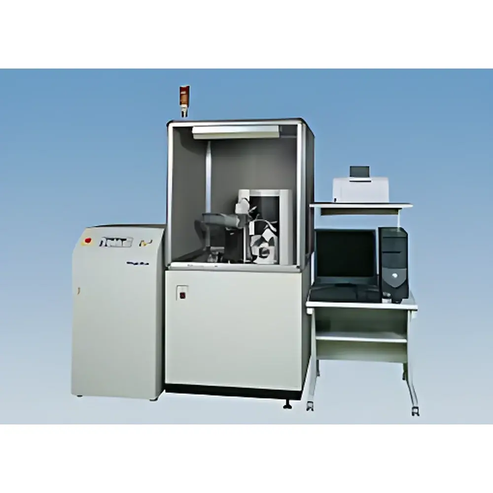
Rigaku D/max RAPID Micro-Area X-ray Diffractometer
| Brand | Rigaku |
|---|---|
| Origin | Japan |
| Model | D/max RAPID |
| Instrument Type | Powder X-ray Diffractometer |
| Configuration | Floor-Standing |
| X-ray Source Options | 3 kW Sealed-Tube or 18 kW Rotating-Anode Anode |
| Detector | Large-Area Cylindrical 2D Imaging Plate (IP), 466 mm × 256 mm |
| Angular Range | –60° to +144° 2θ |
| Beam Conditioning | Incident-Beam Monochromator |
| Collimators | 800, 300, 100, 50, 30 µm (optional 10 µm) |
| Compliance | Designed for ISO/IEC 17025-compliant laboratories |
Overview
The Rigaku D/max RAPID Micro-Area X-ray Diffractometer is a floor-standing, high-flexibility powder X-ray diffractometer engineered for structural characterization of spatially constrained or physically delicate samples. It operates on the Bragg diffraction principle using Cu Kα radiation (λ = 1.5418 Å), with optional monochromatic beam conditioning to suppress Kβ emission and enhance peak resolution. Unlike conventional θ–2θ scanning systems, the D/max RAPID employs a large-area cylindrical imaging plate (IP) detector—466 mm × 256 mm active area—capable of capturing diffraction data over an extended angular range from –60° to +144° in 2θ in a single exposure. This geometry enables rapid, non-destructive acquisition of Debye–Scherrer rings without mechanical goniometer movement, significantly reducing measurement time and mechanical drift artifacts. The system supports both sealed-tube (3 kW) and high-power rotating-anode (18 kW) X-ray sources, allowing users to balance intensity requirements against thermal load management and operational longevity.
Key Features
- Micro-area analysis capability enabled by interchangeable precision collimators (30–800 µm diameter, with optional 10 µm); ideal for mapping heterogeneous samples or isolating specific microstructural domains.
- Dedicated high-resolution CCD camera integrated into the goniometer head for real-time, parfocal sample visualization—critical for precise positioning of irregular, non-pelletized, or liquid-containing specimens.
- Incident-beam monochromator ensures high spectral purity, minimizing background scatter and improving signal-to-noise ratio for weak diffraction signals from thin films or trace phases.
- Robust floor-standing architecture with vibration-damped optical bench, optimized for long-term stability during extended acquisitions such as in situ heating experiments or time-resolved studies.
- Modular design accommodates a full suite of accessories—including high-temperature stages (up to 1200 °C), humidity-controlled cells, and fiber texture holders—without compromising alignment integrity.
Sample Compatibility & Compliance
The D/max RAPID excels in analyzing samples that cannot be prepared as conventional pressed pellets: free-standing thin films, microliter-volume liquid suspensions, irregular particulates, embedded cross-sections, and fiber bundles. Its low-divergence microbeam configuration maintains sufficient flux even at sub-100 µm spot sizes, enabling phase identification in regions < 0.01 mm². The instrument meets mechanical and electrical safety standards per IEC 61010-1 and is fully compatible with laboratory quality management frameworks. Data acquisition software supports user-defined metadata tagging, electronic signatures, and export formats compliant with ASTM E1421 and ISO 17873 for XRD data reporting. When deployed in regulated environments, the system can be validated per IQ/OQ protocols aligned with FDA 21 CFR Part 11 requirements for electronic records and signatures.
Software & Data Management
Acquisition and analysis are managed through Rigaku’s proprietary SmartLab Studio II platform, which provides automated calibration, real-time ring integration, and quantitative phase analysis (QPA) via Rietveld refinement (using PDXL or TOPAS engines). Raw 2D images are stored in TIFF or binary formats with embedded header metadata (wavelength, collimator ID, exposure time, sample position). The software supports batch processing across multiple sample positions, enabling automated micro-diffraction mapping. Audit trail functionality logs all parameter changes, user actions, and processing steps—essential for GLP audits and method transfer between sites. Export options include CIF, XYE, and XML-based XRD-ML for interoperability with third-party crystallographic databases and LIMS integrations.
Applications
- Powder phase identification and quantification: Rapid screening of polymorphic forms, impurity detection, and crystallinity assessment in pharmaceuticals and battery cathode materials.
- Thin-film phase and orientation analysis: Determination of preferred orientation (texture), epitaxial relationships, and interfacial strain in sputtered or CVD-grown layers.
- Residual stress evaluation: Sin²ψ method implementation using asymmetric reflections to quantify macroscopic stress states in coated metals and ceramics.
- Fiber diffraction and crystallite size distribution: Analysis of polymer fibers, natural biopolymers, and nanocellulose assemblies using azimuthal integration and Scherrer analysis.
- In situ and operando studies: Real-time monitoring of phase transformations during controlled heating (e.g., calcination, decomposition) or gas exposure, leveraging the system’s fast 2D acquisition speed.
FAQ
What sample preparation methods are recommended for micro-area XRD on the D/max RAPID?
Minimal preparation is required: flat, stable mounting is sufficient. Free-standing films may be affixed to Kapton tape; powders can be loaded directly into capillaries or onto low-background silicon wafers.
Can the D/max RAPID perform Rietveld refinement?
Yes—integrated SmartLab Studio II supports full-pattern Rietveld analysis when coupled with PDXL or external TOPAS licenses.
Is the system compatible with automated sample changers?
While not standard, the goniometer design permits third-party robotic integration under custom engineering support from Rigaku Applications Group.
What is the typical measurement time for a single micro-area diffraction pattern?
With a 30 µm beam and 3 kW source, acquisition times range from 30 seconds to 5 minutes depending on sample scattering power and desired signal-to-noise ratio.
Does the imaging plate detector require periodic recalibration?
Yes—detector response uniformity and spatial distortion correction are performed during initial installation and recommended annually or after major maintenance events.
")


")