Montana Instruments CryoRAMAN Ultra-High-Resolution Low-Temperature Micro-Raman System
| Brand | Montana Instruments |
|---|---|
| Origin | USA |
| Model | CryoRAMAN |
| Instrument Type | Grating-Based Raman Spectrometer |
| Spectral Range | 190 nm – mid-IR (down to 150 nm with custom coatings, gratings & detectors) |
| Spectral Resolution | 0.08 nm (CCD), 0.05 nm (PMT) across entire focal plane |
| Spatial Resolution | ≥15 lp/mm @ 50% modulation (center of focal plane) |
| Minimum Raman Shift | 0.8 cm⁻¹ |
| Temperature Range | 4 K – 350 K (600 K optional) |
| Sample Positional Drift | <20 µm (4.2 K–350 K, optical axis) |
| Excitation Wavelengths | 532 nm (standard), 785 nm (optional), customizable UV–NIR–MIR sources |
| Laser Spot Size | 1–3 µm |
| Field of View | >30 µm |
| Objective NA | 0.75 (Cryo-Optic®) |
| Spectrometer Options | FERGIE® (f/4, 80.08 mm FL) or IsoPlane® SCT320 (f/4.6, 320 mm FL) |
| Polarization-Resolved Capability | Yes (interchangeable polarization filtering modules) |
| Electrical Transport Integration | CryoChip16-compatible, 16-channel DC measurement platform |
| Automation | Fully automated cryostation (Cryostation® S100), touch-screen + PC control |
| Compliance | Designed for GLP/GMP-aligned workflows |
Overview
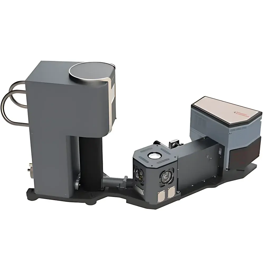
The Montana Instruments CryoRAMAN Ultra-High-Resolution Low-Temperature Micro-Raman System is a fully integrated, research-grade platform engineered for quantitative, spatially resolved vibrational spectroscopy under precisely controlled cryogenic and variable-temperature conditions. Combining Montana Instruments’ proprietary Cryostation® S100 closed-cycle cryogenic platform with Princeton Instruments’ high-fidelity spectroscopic engines—FERGIE® or IsoPlane® SCT320—the system implements a rigid, bridge-coupled optical architecture that eliminates manual alignment while preserving diffraction-limited performance across the full spectral and thermal operating range (4 K to 350 K, extendable to 600 K). Its core measurement principle relies on inelastic laser scattering (Raman effect), enhanced by confocal micro-optics and low-noise, back-illuminated CCD or deep-cooled PMT detection. The system enables direct correlation between structural dynamics (phonon softening, mode splitting, linewidth broadening), electronic transitions (PL quenching, exciton binding energy shifts), and thermally activated transport phenomena—making it indispensable for condensed matter physics, quantum materials science, and nanoscale device characterization.
Key Features
- Automated, helium-free cryogenics: Cryostation® S100 delivers stable base temperatures down to 4 K with <30 s thermal settling per setpoint and <1 µm/°C positional drift across the full 4–350 K range.
- Zero-aberration Cryo-Optic® imaging: High-NA (0.75) objective optimized for cryogenic operation ensures diffraction-limited spot size (1–3 µm), high collection efficiency, and minimal thermal-induced wavefront distortion.
- Dual-spectrometer flexibility: Select FERGIE® for rapid, wide-band (400–1100 nm), low-noise acquisition or IsoPlane® SCT320 for ultra-high-resolution (<0.8 cm⁻¹ wavenumber resolution) and zero-astigmatism imaging across extended spectral ranges (190 nm–mid-IR).
- Modular excitation & detection: Interchangeable DPSS lasers (532 nm standard; 785 nm, 633 nm, or custom wavelengths available), polarization-resolved filtering units, and motorized slit selection (10–500 µm) support method development for diverse sample classes.
- Synchronized multimodal acquisition: Simultaneous Raman/PL spectroscopy, white-light microscopy (3 MP camera + tunable HB LED), and DC electrical transport (via CryoChip16 interface) enable correlative structure–function–property analysis without sample repositioning.
- Touchscreen + software-controlled operation: Intuitive GUI supports automated temperature ramps, spectral mapping (XY/Z raster), and real-time data preview—reducing operator dependency and improving experimental reproducibility.
Sample Compatibility & Compliance
The CryoRAMAN system accommodates bulk crystals, exfoliated 2D flakes (graphene, TMDs), thin films, heterostructures, and biological specimens (e.g., cryo-preserved protein aggregates or lipid bilayers) within a 10 × 10 × 2.5 mm sample chamber (custom mounts available). Its ATSM (Advanced Thermal Stage Module) features low-thermal-mass design and active position stabilization, ensuring sub-micron spatial registration during thermal cycling—a prerequisite for quantitative hyperspectral imaging. All optical components are vacuum-compatible and rated for repeated thermal cycling between 4 K and 350 K. From a regulatory standpoint, the system supports audit-ready workflows: wavelength calibration traceability to NIST-traceable Hg/Ne lamps, mechanical repeatability ≤ ±0.0015 nm post-calibration, and timestamped metadata logging compatible with FDA 21 CFR Part 11–aligned electronic lab notebook (ELN) integration.
Software & Data Management
Acquisition and analysis are managed through Princeton Instruments’ LightField® software, which provides scriptable automation (Python API), real-time spectral fitting (Voigt, Lorentzian, Gaussian deconvolution), and multidimensional data export (HDF5, TIFF, CSV). Raw spectra include embedded metadata: temperature, laser power, integration time, grating position, slit width, and detector gain—all synchronized to millisecond precision. For long-term data integrity, LightField supports automatic checksum generation, version-controlled experiment templates, and secure user-role permissions. Exported datasets conform to FAIR principles (Findable, Accessible, Interoperable, Reusable) and integrate natively with third-party platforms including Igor Pro, MATLAB, and Python-based analysis stacks (e.g., SciPy, scikit-learn for clustering of temperature-dependent mode evolution).
Applications
- Quantum phase transitions: Tracking Mott insulator–metal crossover in RuCl₃ or CrCl₃ via phonon collapse and linewidth divergence below critical temperature.
- Low-dimensional phonon engineering: Quantifying anharmonic decay rates in monolayer MoS₂ through temperature-dependent Raman shift (dω/dT) and FWHM analysis from 5 K to 300 K.
- Strain–temperature coupling: Mapping local lattice expansion in twisted bilayer graphene using spatially resolved 2D-mode splitting under controlled thermal gradients.
- Exciton thermodynamics: Correlating PL peak energy, linewidth, and intensity with carrier thermalization pathways in WSe₂ heterobilayers.
- Cryobiophysics: Probing hydration-shell dynamics and secondary-structure stability in amyloid fibrils via amide I band shifts under sub-100 K conditions.
- Device-level metrology: In situ Raman–IV characterization of van der Waals transistors to identify charge-transfer-induced mode suppression and contact resistance evolution.
FAQ
What is the lowest achievable temperature, and does the system require liquid helium?
The CryoRAMAN operates down to 4 K using a fully automated, closed-cycle Cryostation® S100—no liquid helium, no cryogen handling, and no specialized cryogenic training required.
Can I perform polarization-resolved Raman measurements?
Yes. Optional motorized polarization analyzers and rotatable half-wave plates integrate seamlessly into the excitation and collection paths, enabling full Stokes vector acquisition for symmetry analysis of crystalline and layered materials.
Is electrical transport measurement truly simultaneous with Raman acquisition?
Yes. The CryoChip16 stage provides 16 independent DC channels with sub-picoamp current sensitivity, and all signals—including spectrometer triggers, temperature logs, and voltage sweeps—are hardware-synchronized via TTL pulses and shared clock distribution.
How is spectral calibration maintained across temperature cycles?
Each acquisition includes on-the-fly reference lamp exposure (Hg/Ne), and LightField applies pixel-wise nonlinearity correction based on pre-characterized grating dispersion models validated at multiple temperatures (4 K, 77 K, 300 K).
What level of spatial registration accuracy is achieved during thermal ramping?
ATSM’s low-drift mechanics limit sample displacement to <100 nm peak-to-peak under isothermal conditions and <1 µm/°C across the full 4–350 K range—enabling reliable 2D Raman mapping over multi-hour temperature sweeps.