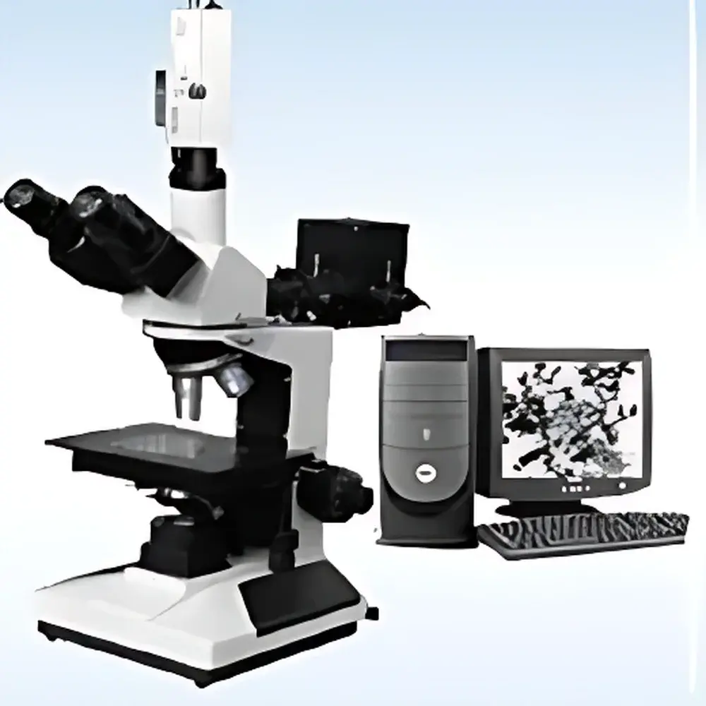
EMCN MDM-300 Upright Reflected & Transmitted Light Metallurgical Microscope
| Brand | EMCN |
|---|---|
| Model | MDM-300 |
| Configuration | Upright, Trinocular |
| Total Magnification Range | 50×–600× |
| Eyepieces | Wide-Field WF10× (Φ18 mm) |
| Objectives | PL 5×/0.12 (WD 18.3 mm), PLL 10×/0.25 (WD 8.9 mm), PLL 40×/0.60 (spring-loaded, WD 3.7 mm), PL 60×/0.85 (spring-loaded, WD 0.26 mm) |
| Focusing Mechanism | Coaxial coarse/fine focus with 15 mm travel |
| fine adjustment resolution | 2 µm |
| Illumination | Dual-path halogen-based system — reflected (epi-illumination) with polarizing filter and adjustable brightness |
| Polarization | Insertable linear polarizer and analyzer (rotatable analyzer in trinocular tube) |
| Tube Angle | 30° inclined, rotating trinocular head with beam splitter for simultaneous visual observation and camera output |
| Compliance | Designed to meet ISO 8578 (microscopy — terminology and definitions) and ASTM E3 (standard guide for preparation of metallographic specimens) |
Overview
The EMCN MDM-300 is an upright metallurgical microscope engineered for high-fidelity microstructural analysis of both opaque and semi-transparent specimens under reflected (epi-) and transmitted illumination modes. Its dual-illumination architecture enables rigorous metallographic evaluation—such as grain boundary delineation, phase identification, inclusion analysis, and coating thickness assessment—as well as complementary applications in materials science, semiconductor inspection, and advanced ceramics characterization. The instrument operates on standard Köhler illumination principles for both optical paths, ensuring uniform brightness, minimized glare, and optimal contrast across the full magnification range (50×–600×). Its rigid mechanical design features a precision-machined base and coaxial focusing mechanism with 2 µm fine-focus resolution, supporting reproducible Z-axis positioning essential for comparative analysis and serial sectioning studies.
Key Features
- Trinocular observation head with 30° inclination and beam-splitting capability (typically 50:50 or 70:30 split ratio) for concurrent eyepiece viewing and digital image acquisition
- Four-objective parfocal turret with plan achromat (PL/PLL) optics optimized for metallurgical imaging—free of cover-slip correction requirements and corrected for field flatness and chromatic aberration up to 60×
- Dual independent halogen illumination systems: reflected-light path with built-in polarizer and rotatable analyzer (enabling brightfield, darkfield, and polarized contrast modes); transmitted-light path with multi-filter wheel (blue, yellow, green, ground-glass diffuser) and stabilized current supply
- Coaxial coarse/fine focusing with 15 mm vertical travel and calibrated micrometer scale; fine-focus graduation at 2 µm per division ensures precise depth-of-field localization
- Insertable polarization components conforming to ISO 10110-6 standards for stress birefringence evaluation and anisotropic phase discrimination
- Modular platform compatible with industry-standard C-mount adapters (e.g., 1× or 0.5× reduction lenses) for integration with CCD/CMOS cameras, frame grabbers, and A/D imaging systems
Sample Compatibility & Compliance
The MDM-300 accommodates standard 25 mm and 32 mm diameter metallographic mounts, as well as thin-sectioned non-metallic specimens (e.g., geological thin sections, polymer films, and coated substrates) when used in transmitted mode. Its long-working-distance objectives support examination of unpolished or lightly prepared surfaces—including as-cast alloys, fractured specimens, and electroplated layers—without risk of objective contact. The system complies with ASTM E3 (specimen preparation), ASTM E407 (etchant selection), and ISO 643 (steel microstructure rating), and its optical performance aligns with ISO 8578 definitions for resolution, field number, and numerical aperture fidelity. When paired with FDA 21 CFR Part 11–compliant imaging software (e.g., NIS-Elements, Olympus cellSens, or Media Cybernetics Image-Pro), the platform supports audit-trail-enabled documentation required under GLP and GMP environments.
Software & Data Management
While the MDM-300 itself is hardware-only, its trinocular port and standardized C-mount interface enable seamless coupling with third-party digital imaging solutions. Recommended configurations include progressive-scan color CCD cameras (e.g., Sony ICX series sensors) and USB 3.0 or GigE frame grabbers supporting 12-bit dynamic range and real-time histogram monitoring. Compatible software packages provide measurement tools (line, area, particle count), calibration traceability via stage micrometers, and export to TIFF/PNG/DICOM formats. Metadata embedding (objective ID, magnification, illumination mode, exposure time) ensures data integrity during regulatory review. Optional motorized Z-drive and XY stage integration further enable automated tile scanning and focus stacking—critical for quantitative stereology and ASTM E112 grain size analysis.
Applications
- Metallography: Grain size determination (ASTM E112), inclusion rating (ASTM E45), decarburization depth measurement, weld zone analysis
- Electronics: PCB trace integrity, solder joint void analysis, IC package delamination screening
- Advanced Materials: Ceramic grain morphology, composite fiber distribution, thermal barrier coating porosity quantification
- Forensics & Failure Analysis: Fractography, toolmark comparison, paint layer stratigraphy
- Academic Research: Phase transformation kinetics, recrystallization behavior, corrosion product identification via polarized light contrast
FAQ
Does the MDM-300 support DIC (Differential Interference Contrast)?
No—the MDM-300 does not include Wollaston prisms or Nomarski optics; it is configured for brightfield, darkfield, and linear polarized contrast only.
Can I use oil immersion objectives with this microscope?
Not without modification—the standard objective set is dry-type only; oil immersion objectives require matching condenser NA and immersion medium compatibility not provided in the base configuration.
Is the transmitted light path suitable for biological tissue sections?
Yes, provided specimens are mounted on standard glass slides and coverslips; however, the system lacks condenser height adjustment and centering controls typical of dedicated biological microscopes.
What is the maximum specimen height clearance under the objectives?
Approximately 28 mm beneath the 5× objective (WD 18.3 mm) and ≥5 mm beneath the 60× objective (WD 0.26 mm), accommodating most standard metallographic mounts and low-profile sample holders.
How is calibration traceability maintained for quantitative measurements?
Calibration relies on NIST-traceable stage micrometers and certified graticules; users must perform periodic verification per ISO/IEC 17025 guidelines when operating in regulated quality assurance settings.


