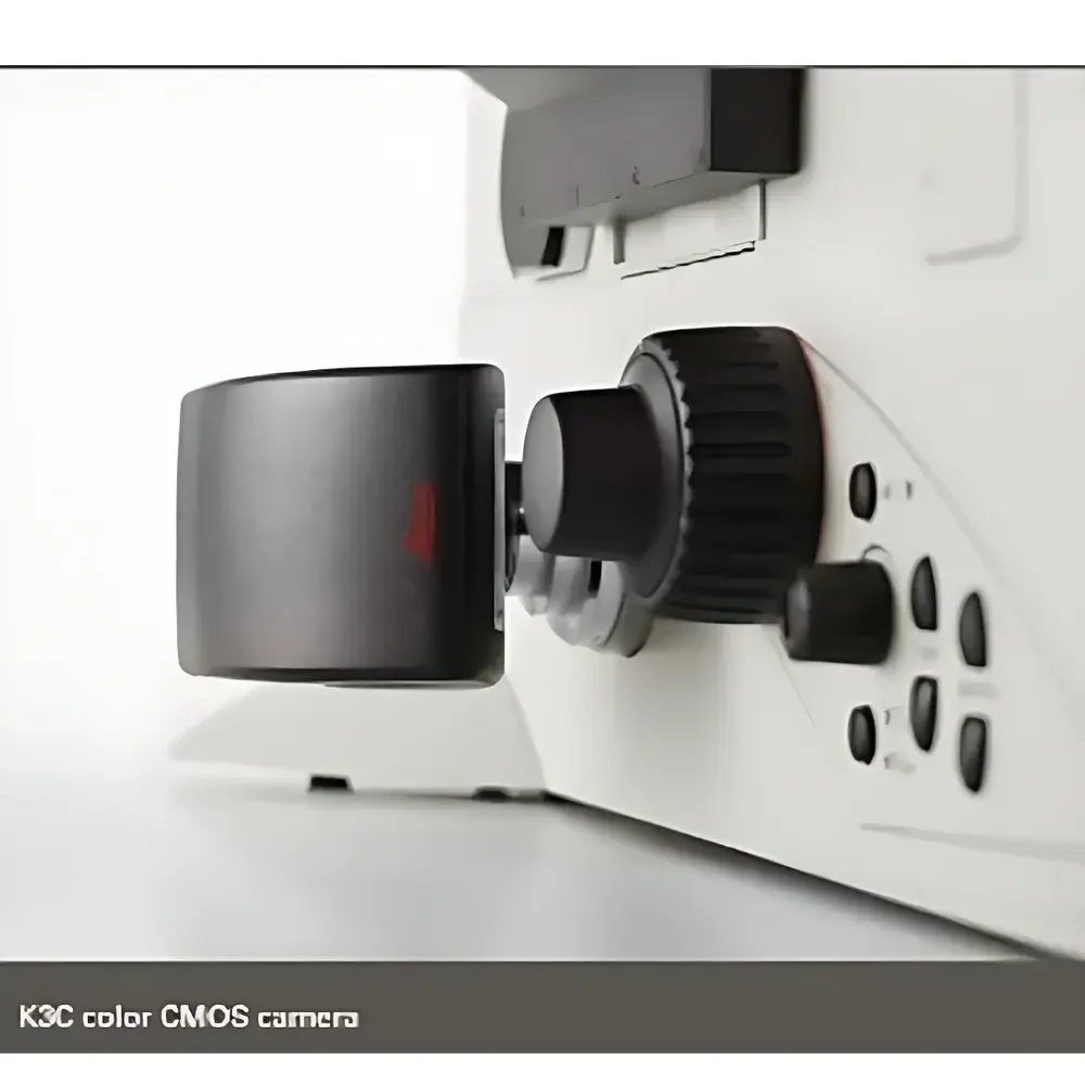
Leica K3C & K3M Microscope CMOS Cameras
| Brand | Leica |
|---|---|
| Origin | Germany |
| Model | K3C & K3M |
| Sensor Type | CMOS |
| Color Capability | K3C (Color), K3M (Monochrome) |
| Bit Depth | 12-bit per channel (RGB for K3C) |
| Dynamic Range | High |
| Frame Rate | High-speed acquisition |
| Interface | USB 3.0 |
| Compliance | CE, RoHS, ISO 9001 certified manufacturing |
| Intended Use | Integrated microscopy imaging for life science, clinical diagnostics, industrial QA/QC, and forensic analysis |
Overview
The Leica K3C and K3M are high-performance, USB 3.0–enabled CMOS microscope cameras engineered for precision digital imaging across demanding scientific and industrial workflows. Designed as native imaging partners for Leica Microsystems’ upright, inverted, and stereo microscopes, these cameras operate on a shared hardware platform optimized for low-noise signal acquisition, wide dynamic range, and deterministic timing—critical attributes for quantitative brightfield, phase contrast, DIC, and fluorescence microscopy. The K3C employs a color CMOS sensor with true 12-bit RGB digitization per channel, enabling accurate spectral fidelity and reproducible colorimetric analysis without interpolation artifacts. The K3M utilizes an identical optical path and electronics architecture but features a monochrome sensor, delivering higher quantum efficiency and superior signal-to-noise ratio (SNR) for low-light applications such as weak fluorescence, FISH, or time-lapse imaging where photon budget is constrained. Both models implement hardware-level binning, region-of-interest (ROI) readout, and global shutter functionality to eliminate motion blur during rapid stage scanning or automated tile acquisition.
Key Features
- High-sensitivity CMOS sensors with back-illuminated architecture (K3M) or advanced color filter array (K3C), optimized for >75% peak QE in visible spectrum
- True 12-bit linear digitization per RGB channel (K3C) or per grayscale channel (K3M), supporting calibrated intensity measurements and downstream pixel-based quantification
- Dynamic range exceeding 70 dB (typical), enabling simultaneous capture of saturated and sub-threshold signal regions within single exposures—essential for heterogeneous fluorescent samples or reflective metallurgical surfaces
- USB 3.0 interface with sustained data throughput up to 400 MB/s, facilitating full-frame acquisition at >30 fps (2048 × 1536) and accelerated tile-based mosaicking
- On-camera hardware features including programmable exposure control, gain adjustment, black level calibration, and temperature-stabilized sensor housing for thermal noise suppression
- Zero-latency trigger synchronization via TTL input/output for integration into automated microscopy platforms and multi-modal imaging rigs
Sample Compatibility & Compliance
The K3C and K3M support standardized sample types across regulated and research environments: fixed and live fluorescently labeled cells and tissues (e.g., immunofluorescence, GFP/RFP transfection), histopathological sections, semiconductor wafers, metallographic specimens (steel, aluminum alloys), particulate contamination samples (ISO 16232, VDA 19), and forensic trace evidence (hair, fibers, gunshot residue). Both cameras comply with IEC 61000-6-3 (EMC emission standards), carry CE marking under the EU Medical Device Regulation (MDR) Annex XVI for non-invasive diagnostic imaging, and conform to RoHS 2011/65/EU. Firmware and driver stacks are validated for GLP/GMP-aligned laboratories, supporting audit trails and user-access controls when deployed with Leica Application Suite X (LAS X) software under FDA 21 CFR Part 11-compliant configuration.
Software & Data Management
Native integration with Leica LAS X software provides full camera control, real-time image optimization, multi-channel fluorescence alignment, Z-stack reconstruction, and automated mosaic stitching with overlap-based registration and intensity normalization. Raw image data is saved in TIFF format with embedded EXIF metadata (exposure time, gain, lens ID, objective magnification, calibration timestamp). LAS X supports DICOM export for PACS integration in clinical pathology settings and HDF5 container format for high-throughput computational pipelines. The SDK (Software Development Kit) enables custom integration with Python (via PyLeica), MATLAB, or LabVIEW for algorithm-driven analysis, machine learning preprocessing, or LIMS interfacing. All firmware updates undergo version-controlled release validation and include rollback capability.
Applications
- Life sciences: Quantitative cell morphology analysis, mitotic index scoring, co-localization studies, and time-lapse tracking of organelle dynamics
- Clinical pathology: Digital slide scanning support for frozen section review, immunohistochemistry (IHC) scoring, and cytology screening workflows
- Industrial quality assurance: Surface defect detection on PCBs, cleanliness verification per ISO 16232 Class C–F, grain structure evaluation in heat-treated alloys
- Forensic science: High-fidelity documentation of toolmarks, fiber comparison, and trace evidence photomicrography compliant with SWGMAT guidelines
- Materials science: In-situ corrosion monitoring, coating thickness measurement via interference contrast, and particle size distribution analysis using calibrated pixel-to-micron mapping
FAQ
What is the difference between the K3C and K3M in terms of sensor performance?
The K3C uses a color CMOS sensor with Bayer-pattern filtering and delivers 12-bit RGB output; the K3M uses a monochrome sensor with no color filter, achieving ~30% higher quantum efficiency and lower read noise—ideal for low-signal fluorescence or quantitative grayscale applications.
Can the K3C/K3M be used with non-Leica microscopes?
Yes—via C-mount adapter (standard 23.2 mm flange distance) and third-party drivers compatible with GenICam-compliant USB3 Vision protocols; however, full feature access (e.g., hardware-triggered Z-stacking, objective-linked magnification scaling) requires Leica hardware integration.
Is the camera suitable for GxP-regulated environments?
When deployed with LAS X in validated configuration—including electronic signatures, change control logs, and audit trail activation—the system meets requirements for FDA 21 CFR Part 11, EU Annex 11, and ISO/IEC 17025 documentation integrity standards.
How does the dynamic range benefit fluorescence imaging?
A high dynamic range allows single-exposure capture of both dim cytoplasmic signals and saturated nuclear markers—eliminating the need for exposure bracketing and reducing phototoxicity during live-cell imaging.
What calibration options are available for quantitative intensity measurements?
Leica provides NIST-traceable flat-field and dark-frame calibration kits; users can perform daily offset/gain correction and apply pixel-wise sensitivity maps directly within LAS X for absolute intensity normalization across sessions and instruments.


