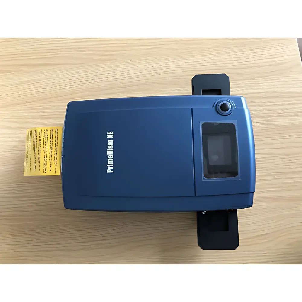
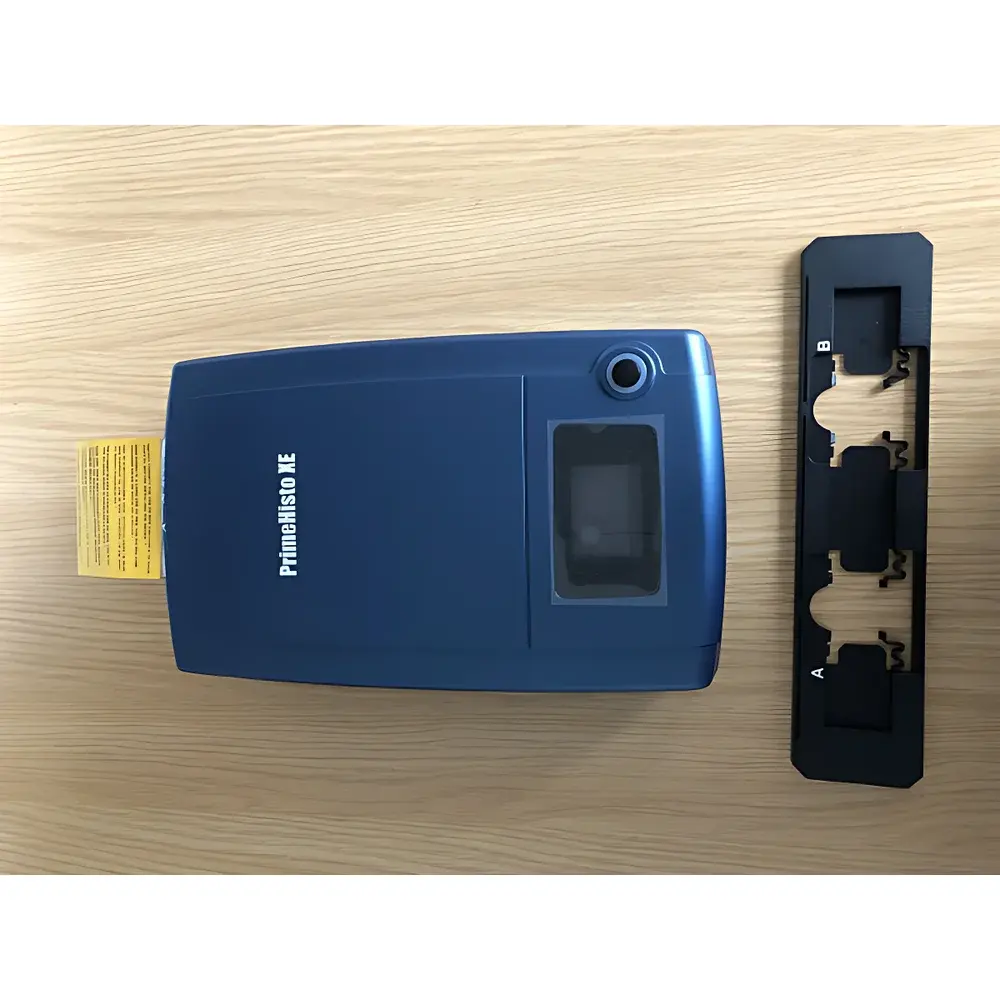
Truelab PrimeHisto XE Whole-Slide Digital Scanner
| Brand | Truelab |
|---|---|
| Model | PrimeHisto XE |
| Origin | Shanghai, China |
| Slide Format | 25 × 75 mm (1 × 3 in) |
| Scan Area | 24.3 × 36.5 mm |
| Optical Resolution | 10,000 dpi |
| Dynamic Range | 3.8 OD |
| Illumination | Transmissive White LED |
| Image Formats | TIFF, JPEG, BMP |
| Dimensions | 260 × 155 × 70 mm |
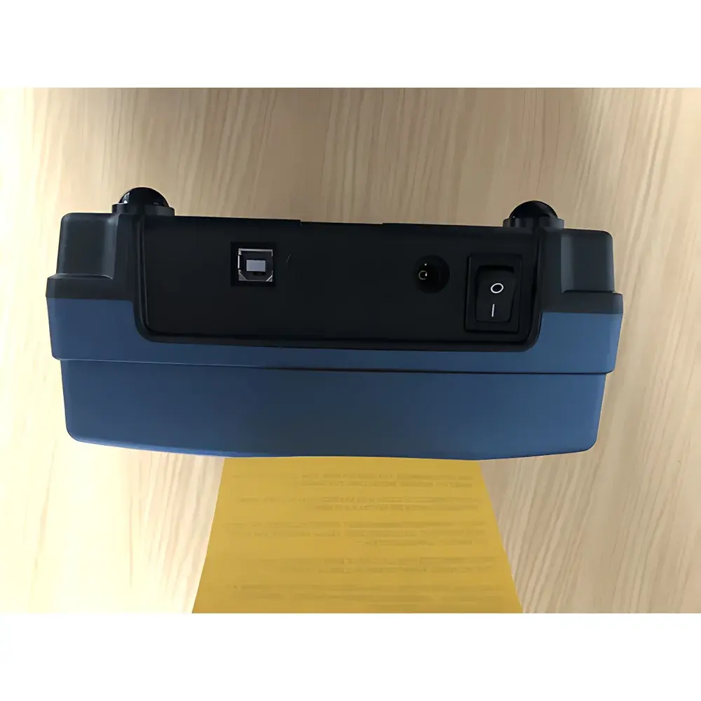
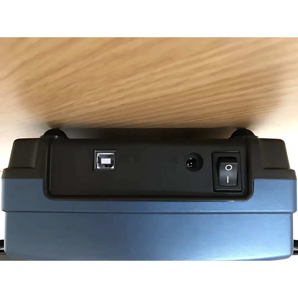
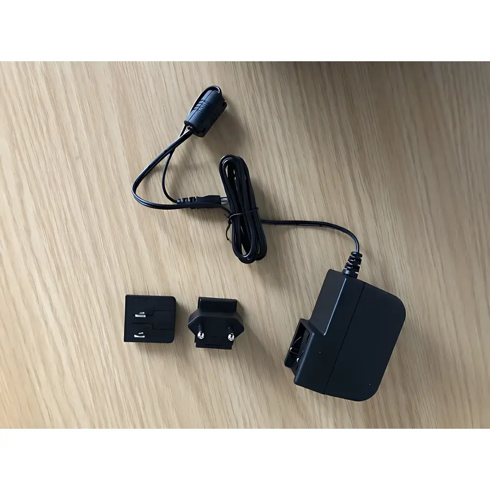
| Power Supply | 100–240 V AC, 12 V / 1.5 A adapter |
| OS Compatibility | Windows XP–10, macOS 10.5+ |
| Software | HistoView v3.x (driver-integrated acquisition & post-processing suite) |
| Optional Accessory | Rotatable Polarizer Mount for geological thin-section analysis |
Overview
The Truelab PrimeHisto XE is a high-fidelity whole-slide digital scanner engineered for routine and research-grade digitization of histological and cytological specimens mounted on standard 25 × 75 mm glass slides. It operates on a transmissive brightfield optical architecture, utilizing a precision-stepped motorized stage and a high-sensitivity monochrome or color CMOS sensor (configurable per system variant) to capture full-slide images at up to 10,000 dpi native optical resolution—equivalent to ~75× magnification in conventional microscopy terms. Unlike slide scanners relying on tiling-based stitching algorithms with inherent parallax or focus drift, the PrimeHisto XE employs a single-pass linear scanning mode with real-time auto-focus feedback and dynamic exposure control, ensuring consistent pixel fidelity across the entire 24.3 × 36.5 mm scan field. Its 3.8 optical density (OD) dynamic range enables reliable visualization of both densely stained nuclei and faint cytoplasmic structures within the same field—critical for morphometric quantification and educational annotation tasks.
Key Features
- Native 10,000 dpi optical resolution with sub-micron pixel pitch (≤0.25 µm/pixel), supporting quantitative morphological analysis without interpolation artifacts
- Transmissive white LED illumination system with uniformity >95% across full scan area, calibrated per ASTM E308-22 spectral standards
- Integrated real-time autofocus loop using contrast-detection algorithm, optimized for variable tissue thickness (3–15 µm typical)
- HistoView software v3.x—FDA 21 CFR Part 11–compliant audit trail module available as optional license; supports DICOM-SR export for PACS integration
- Compact benchtop footprint (260 × 155 × 70 mm) with low-power consumption (<18 W idle, <25 W active), suitable for shared lab environments and teaching facilities
- Optional rotatable polarizer mount (sold separately) enabling dual-mode acquisition: standard brightfield + compensated polarized light for birefringent material identification
Sample Compatibility & Compliance
The PrimeHisto XE accommodates all standard histopathology slides prepared using conventional protocols—including hematoxylin & eosin (H&E), immunohistochemistry (IHC), special stains (e.g., Masson’s trichrome, PAS), and fluorescence-labeled sections (when equipped with appropriate excitation filters). It complies with ISO 15189:2022 requirements for pre-analytical instrumentation used in accredited medical laboratories. For geological and materials science applications, the optional polarizer mount permits standardized analysis of polished thin sections per ASTM D5777 (mineral identification) and ISO 14488 (fiber morphology assessment). All firmware and HistoView software versions undergo annual GLP-aligned validation per internal SOP-IM-027, with traceable calibration certificates issued for each unit prior to shipment.
Software & Data Management
HistoView serves as both acquisition engine and image management platform. It provides lossless TIFF export (16-bit grayscale or 24-bit RGB), JPEG compression with adjustable quality factor (Q=85–100), and batch metadata tagging (slide ID, date/time stamp, operator ID, staining protocol). The software supports region-of-interest (ROI) annotation, multi-layer overlay (e.g., nuclear mask + membrane stain), and basic measurement tools (area, perimeter, intensity histogram). For regulated environments, the Part 11–enabled version includes electronic signatures, session lockout, and immutable audit logs stored in SQLite3 database with SHA-256 hash verification. Export formats include OME-TIFF for interoperability with QuPath, HALO, and OpenSlide-compatible viewers.
Applications
- Clinical pathology: Digital archiving of surgical pathology slides for second-opinion consultation and telepathology workflows
- Academic research: Longitudinal tissue morphology tracking in rodent models, tumor microenvironment mapping, and spatial transcriptomics correlation
- Education: High-resolution slide libraries for undergraduate histology labs, virtual microscopy exams compliant with AAMC guidelines
- Geosciences: Quantitative mineral phase identification in petroleum exploration cores and metamorphic rock thin sections
- Materials science: Birefringence characterization of polymer blends, liquid crystal domains, and semiconductor wafer defects
- Pharmaceutical QA: Crystal habit analysis of active pharmaceutical ingredients (APIs) per ICH Q5A and USP
FAQ
Does the PrimeHisto XE support fluorescence scanning?
Yes—when configured with optional LED excitation modules (450 nm, 530 nm, 620 nm) and bandpass emission filters, it captures widefield fluorescence signals compatible with common dyes (DAPI, FITC, TRITC).
Can scanned images be imported into AI-based analysis platforms?
All exported TIFF files are OME-compliant and natively supported by QuPath, CellProfiler, and DeepLabCut via standard OpenSlide API wrappers.
Is remote operation possible over LAN/WAN?
HistoView includes built-in VNC-compatible remote desktop access and RESTful API endpoints (JSON/XML) for integration with LIMS and hospital information systems.
What is the maximum slide throughput per hour?
At 40× equivalent resolution (2,500 dpi), average scan time is 42 seconds per slide; throughput exceeds 85 slides/hour in unattended batch mode.
Does the polarizer mount require hardware modification?
No—it attaches magnetically to the existing optical path housing and requires no recalibration; rotation angle is encoded and logged in image metadata.


