MegaSPIM Large-Scale Tissue & High-Throughput Light Sheet Microscope
| Brand | Lifecanvas Technologies |
|---|---|
| Origin | USA |
| Model | MegaSPIM |
| Illumination Geometry | 45° Unilateral Light Sheet Generation with Axial Sweeping (Patent US10989661B2) |
| Sample Chamber Dimensions | >200 mm × 200 mm (X–Y), 50 mm Z-Travel for Specimen Positioning |
| Detection Objective EFL | 180 mm Tube Lens |
| Standard Objective | 3.6×, Large-Field-of-View |
| Optional Objectives | 1.8×, 9×, 15×, 22× |
| Camera | 2048 × 2048 sCMOS, Rolling Shutter Synchronized with Light Sheet Sweep, QE = 82%, Max Frame Rate: 80 fps |
| Dual-Camera Acquisition | Optional, Independently Focused |
| Laser Lines | Standard 3, Expandable to 7 (405–785 nm) |
| Acquisition Workstation | Intel® Core™ i9-12900K (16C/24T), 128 GB Quad-Channel DDR5 RAM, 32 TB Storage (4 × 8 TB Samsung 870 QVO SATA III SSD), Windows 10 Pro, 4K Display |
| Software | MegaSPIM Acquisition & Post-Processing Suite (Real-Time Tile Correction, Stripe Artifact Suppression, GPU-Accelerated 3D Volume Reconstruction, Block-Based Registration) |
| Analysis Workstation (Optional) | AMD Ryzen™ Threadripper PRO 3995WX (64C/128T), 512 GB Quad-Channel DDR4 RAM, 32 TB RAID 0 (4 × 8 TB 7200 RPM HDD), Windows 10 Pro |
Overview
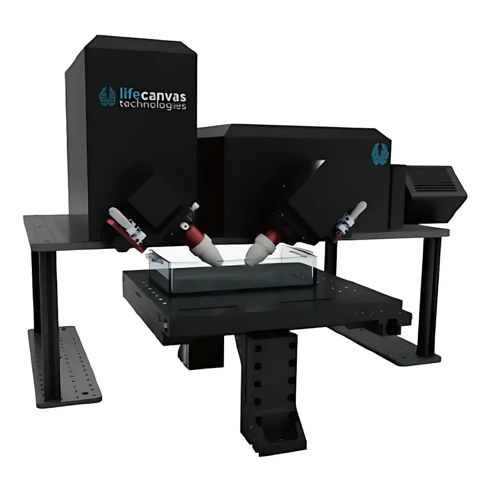
The MegaSPIM Large-Scale Tissue & High-Throughput Light Sheet Microscope is an engineered platform for volumetric fluorescence imaging of centimeter-scale biological specimens with subcellular resolution and minimal phototoxicity. Based on the patented axial light sheet sweeping architecture (US10989661B2), the system generates a thin, dynamically scanned illumination plane at a fixed 45° incidence angle—eliminating static light sheet artifacts while enabling uniform optical sectioning across extended axial ranges. Unlike conventional orthogonal or dual-sided light sheet configurations, the unilateral illumination design simplifies optical alignment, improves compatibility with opaque or scattering samples (e.g., cleared whole organs, embryonic tissue blocks, or organotypic slices), and supports rapid repositioning via motorized Z-stage translation (±50 mm). The system integrates a high-numerical-aperture 180 mm effective focal length (EFL) tube lens with a standard 3.6× large-field objective, delivering a native field of view exceeding 20 mm × 20 mm at the sample plane—ideal for capturing intact murine brains, human cortical biopsies, or developmental zebrafish embryos without tiling. Its modular optical path accommodates up to seven independently controllable laser lines (405–785 nm), ensuring flexibility for multicolor multiplexed labeling strategies in translational histology and spatial omics workflows.
Key Features
- Patented axial light sheet sweeping (US10989661B2) enabling uniform illumination depth penetration and reduced shadowing in thick, heterogeneous tissues
- Large-format sample chamber (>200 mm × 200 mm X–Y footprint) with precision motorized Z-stage (50 mm travel) for multi-position acquisition and serial sectioning alignment
- Standard 3.6× large-FOV objective paired with 180 mm EFL tube lens; optional magnification range from 1.8× (ultra-wide field) to 22× (high-resolution subcellular imaging)
- 2048 × 2048 sCMOS camera with 82% quantum efficiency, rolling shutter synchronized to light sheet sweep timing, supporting up to 80 fps at full resolution
- Dual-camera configuration available with independent focus control—enabling simultaneous orthogonal detection channels or differential refraction correction
- Seven-channel laser combiner (405–785 nm) with individual AOTF or acousto-optic modulation for precise intensity and spectral control per channel
- Acquisition workstation built around Intel Core i9-12900K (16-core), 128 GB quad-channel memory, and 32 TB SATA SSD storage optimized for real-time streaming and buffer management
Sample Compatibility & Compliance
The MegaSPIM platform is validated for imaging optically cleared tissues—including those processed via CLARITY, iDISCO, CUBIC, and SHIELD protocols—as well as live ex vivo preparations such as perfused organ explants and thick brain slices (up to 5 mm). Its open mechanical design permits integration with custom sample holders, perfusion chambers, and environmental control units (temperature, CO₂, humidity). All hardware and software components comply with IEC 61000-6-3 (EMC emissions) and IEC 61000-6-2 (immunity) standards. The acquisition software supports audit trail logging, user access controls, and electronic signature functionality—facilitating alignment with GLP and 21 CFR Part 11 requirements for regulated preclinical research environments.
Software & Data Management
The proprietary MegaSPIM Acquisition & Post-Processing Suite provides a unified interface for instrument control, real-time visualization, and computational reconstruction. Key modules include: (i) tile-based adaptive illumination calibration to correct for beam profile nonuniformity; (ii) GPU-accelerated stripe artifact suppression using Fourier-domain filtering and phase-retrieval algorithms; (iii) block-wise affine and nonrigid registration for stitching multi-position datasets; and (iv) export pipelines compliant with OME-TIFF, N5, and HDF5 formats for downstream analysis in Fiji, Imaris, or custom Python-based workflows. Optional analysis workstations (AMD Threadripper PRO 3995WX, 64-core, 512 GB RAM, RAID 0 HDD array) support batch processing of terabyte-scale volumes—including deconvolution, cell segmentation, and spatial transcriptomics co-registration.
Applications
- Whole-organ 3D mapping of neuronal connectivity in cleared mouse and non-human primate brains
- Longitudinal developmental imaging of embryonic organogenesis in zebrafish and chick models
- Quantitative assessment of tumor microenvironment architecture in human FFPE-derived cleared biopsies
- High-throughput screening of drug-induced structural phenotypes in organoid arrays
- Correlative light-sheet and serial two-photon tomography (SLaM) workflows for multimodal validation
- Spatial proteomics and in situ sequencing data anchoring via fiducial-based volume registration
FAQ
What sample clearing methods are compatible with the MegaSPIM platform?
The system has been empirically validated with aqueous (CLARITY, CUBIC), organic (iDISCO+, uDISCO), and hybrid (SHIELD, PEGASOS) clearing protocols. Refractive index matching between sample and immersion medium is critical; recommended immersion media include FocusClear, RIMS, and 80% glycerol.
Can the MegaSPIM acquire time-lapse data from live specimens?
Yes—when integrated with environmental control units (temperature, gas, humidity), the system supports long-term (≥24 h) imaging of explanted tissues and thick organoids. Low-light sCMOS sensitivity and swept light sheet geometry minimize cumulative photodamage.
Is raw data format compatible with open-source analysis tools?
All acquisitions are saved in OME-TIFF format with embedded metadata (channel wavelengths, exposure times, stage positions, objective parameters), ensuring native compatibility with Fiji/ImageJ, Napari, and the BigDataViewer ecosystem.
Does the software support automated image quality assessment during acquisition?
Yes—the acquisition suite includes real-time metrics for signal-to-noise ratio (SNR), contrast-to-noise ratio (CNR), and focus sharpness (via Laplacian variance), with configurable thresholds that trigger alerts or pause acquisition if degradation exceeds defined limits.
Are service contracts and remote diagnostics available?
Lifecanvas Technologies offers tiered support plans including on-site preventive maintenance, remote firmware/software updates, and priority response SLAs for academic core facilities and industrial R&D laboratories.


