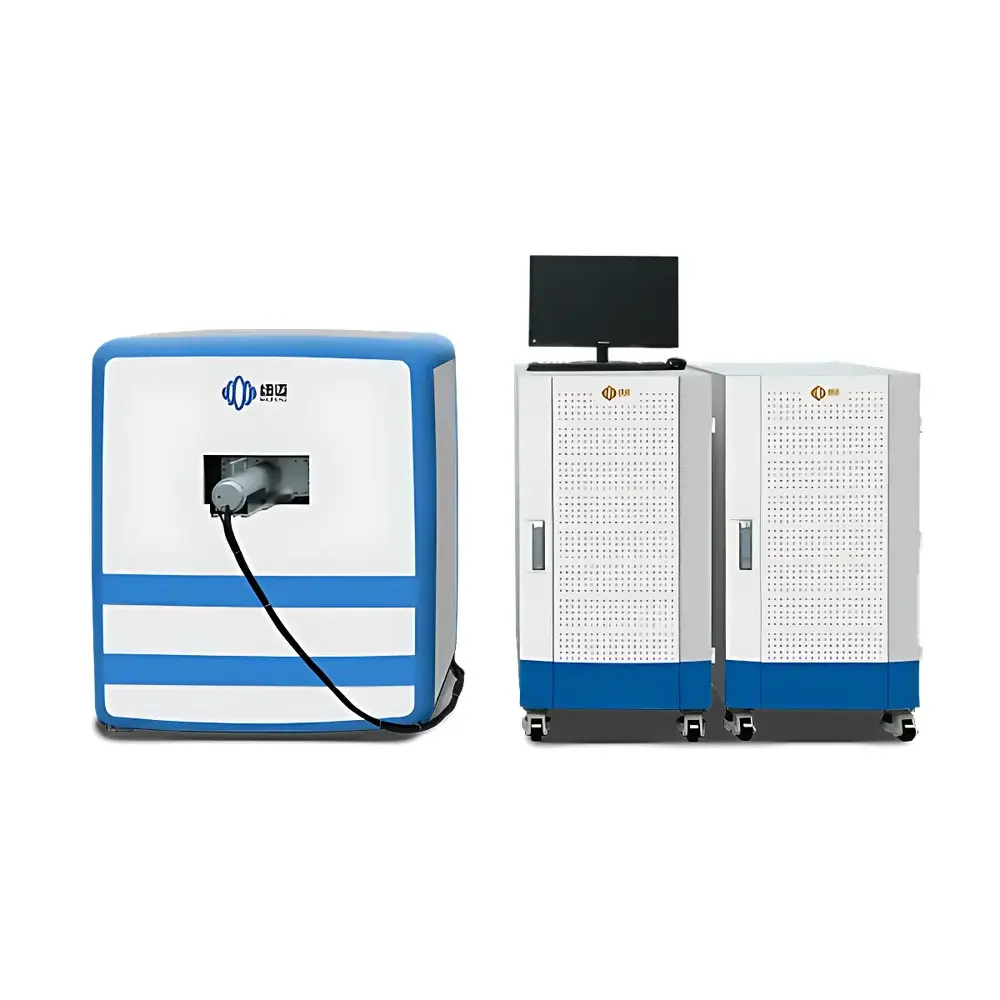
NIUMAG NM42-060H-I Low-Field Nuclear Magnetic Resonance Small Animal Imaging System
| Brand | NIUMAG |
|---|---|
| Origin | Jiangsu, China |
| Manufacturer Type | Authorized Distributor |
| Country of Origin | China |
| Model | NM42-060H-I-1 |
| Instrument Type | Low-Field NMR Imager |
| Sample Compatibility | Solid and Liquid Samples (in vivo rodents, ex vivo tissues, contrast agents, magnetic nanoparticles) |
| Magnet Type | Permanent Magnet |
| Static Field Strength | 1.0 T ± 0.05 T |
| Magnetic Field Homogeneity | ≤30 ppm over 60 mm DSV |
| Sample Weight Range | 1–350 g (mice, rats, excised tissues) |
Overview
The NIUMAG NM42-060H-I is a dedicated low-field nuclear magnetic resonance (NMR) small animal imaging system engineered for preclinical longitudinal studies in biomedical research. Unlike high-field MRI systems requiring cryogenic cooling and RF-shielded rooms, this instrument leverages a permanent magnet architecture to deliver stable, homogeneous static magnetic fields (1.0 T ± 0.05 T) with homogeneity ≤30 ppm over a 60 mm diameter spherical volume (DSV). Its design adheres to core NMR physics principles—specifically, spin-lattice (T1) and spin-spin (T2) relaxation time mapping—and enables non-invasive, label-free, three-dimensional anatomical and functional assessment of live rodents without ionizing radiation. The system supports quantitative analysis of tissue microstructure, edema, necrosis, fat infiltration, and dynamic physiological changes—making it particularly suited for oncology, neurodegenerative disease modeling, metabolic phenotyping, and therapeutic monitoring in mice and rats.
Key Features
- Permanent magnet configuration eliminates dependency on liquid helium, nitrogen, or external power-intensive cooling infrastructure—reducing operational overhead and enabling deployment in standard laboratory environments.
- Integrated gradient coil set with programmable amplitude and slew rate supports multi-contrast pulse sequences including spin-echo, inversion-recovery, and fast spin-echo acquisitions.
- High signal-to-noise ratio (SNR) optimized RF probe tuned for 42.5 MHz (¹H Larmor frequency at 1.0 T), compatible with both in vivo whole-body imaging and high-resolution ex vivo tissue scans.
- Automated shimming routines ensure consistent field homogeneity across repeated sessions—critical for longitudinal reproducibility in pharmacokinetic or disease progression studies.
- Modular hardware architecture allows future expansion with optional diffusion-weighted imaging (DWI), magnetic resonance spectroscopy (MRS), or temperature-controlled animal handling units.
Sample Compatibility & Compliance
The NM42-060H-I accommodates a broad spectrum of biological specimens: live anesthetized rodents (1–350 g), excised organs, tissue biopsies, hydrogels, and nanomaterial suspensions—including iron oxide-based contrast agents and theranostic magnetic nanoparticles. Sample positioning is facilitated by a motorized, laser-guided cradle with integrated physiological monitoring interfaces (respiratory gating, temperature feedback). From a regulatory standpoint, the system complies with IEC 61000-6-3 (EMC emissions) and IEC 61000-6-1 (immunity). While not certified as a medical device under FDA 510(k) or CE-IVD, its data output conforms to DICOM 3.0 standards and supports audit-ready metadata tagging for GLP-compliant preclinical workflows. All acquisition protocols are fully scriptable and version-controlled—enabling traceability required under ISO/IEC 17025 and NIH grant reporting guidelines.
Software & Data Management
Control and reconstruction are performed via NIUMAG’s proprietary NMIStudio platform—a Windows-based application built on Qt and CUDA-accelerated libraries. The software provides real-time preview, interactive ROI definition, pixel-wise T1/T2 mapping, and batch processing pipelines exportable as NIfTI or MATLAB .mat files. Raw k-space data is stored in vendor-neutral HDF5 format with embedded acquisition parameters (TR/TE/TI, flip angle, bandwidth, voxel size). Integrated DICOM gateway enables PACS integration for cross-platform image review. Audit trails log all user actions—including sequence modifications, parameter adjustments, and export events—satisfying FDA 21 CFR Part 11 requirements for electronic records when configured with role-based authentication and digital signature modules. Data encryption at rest and TLS 1.2–secured remote access support institutional IT security policies.
Applications
- Oncology: Monitoring tumor volume, necrosis fraction, and vascular permeability via dynamic contrast-enhanced (DCE) MRI and T2*-weighted susceptibility mapping.
- Neuroscience: Quantifying hippocampal atrophy, white matter integrity loss, and cerebral blood volume changes in transgenic Alzheimer’s or stroke models.
- Nanomedicine: Validating r1/r2 relaxivity of novel contrast agents; tracking biodistribution and clearance kinetics of PEGylated iron oxide nanoparticles.
- Metabolic Research: Assessing hepatic steatosis, pancreatic fat deposition, and brown adipose tissue activation using chemical shift–encoded water-fat separation.
- Cardiovascular Studies: Measuring left ventricular ejection fraction, myocardial edema post-ischemia, and infarct size via late gadolinium enhancement analogues.
FAQ
Is the NM42-060H-I suitable for longitudinal studies in awake animals?
No—motion artifact necessitates anesthesia; however, the system supports isoflurane-compatible gas delivery and physiological monitoring to minimize physiological drift across sessions.
Can the system perform diffusion tensor imaging (DTI)?
Yes—when equipped with optional high-performance gradient coils and advanced diffusion encoding sequences, DTI and tractography are feasible within the 60 mm DSV.
What is the minimum voxel resolution achievable in vivo?
Typical in vivo isotropic resolution is 150–200 µm with adequate SNR; ex vivo scans of fixed tissues achieve ≤75 µm with extended averaging.
Does NIUMAG provide protocol development support for custom applications?
Yes—NIUMAG’s Applications Lab offers collaborative method development, including pulse sequence optimization, phantom validation, and SOP documentation aligned with journal publication standards.
How is system calibration maintained over time?
Daily automated QA routines verify field homogeneity, RF gain stability, and gradient linearity; annual recalibration by certified engineers includes shim map verification and probe Q-factor measurement.
Related Products


