NIUMAG M5-Rat Compact Small Animal MRI System for Arthritis Phenotyping
| Brand | NIUMAG |
|---|---|
| Origin | Israel |
| Manufacturer Type | Authorized Distributor |
| Origin Category | Imported |
| Model | M5-Rat |
| Instrument Type | Nuclear Magnetic Resonance (NMR) Imaging System |
| Magnet Type | Permanent Magnet, 1 Tesla |
| Spatial Resolution | ≤100 µm (in-plane, 3D gradient-echo mode) |
| Imaging Modes | T1-weighted, T2-weighted, PD-weighted, 2D/3D spin-echo and gradient-echo sequences |
| Application Focus | In vivo rodent musculoskeletal phenotyping, arthritis progression monitoring, preclinical OA model validation |
Overview
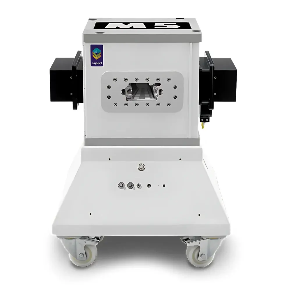
The NIUMAG M5-Rat is a compact, high-field permanent-magnet small animal MRI system engineered specifically for longitudinal in vivo imaging of murine models of osteoarthritis (OA) and other musculoskeletal pathologies. Operating at a stable 1 Tesla field strength, the system leverages robust NMR physics principles—namely proton density contrast, T1 and T2 relaxation time differentiation, and diffusion-weighted signal behavior—to non-invasively visualize articular cartilage degradation, synovial hypertrophy, bone marrow edema, and subchondral sclerosis in live mice and rats. Unlike cryogen-dependent superconducting systems, the M5-Rat employs a self-shielded permanent magnet architecture with near-zero fringe field, enabling safe installation in standard laboratory environments without RF shielding rooms or magnetic safety zones. Its integrated gradient coil set (max. 400 mT/m) and high-sensitivity quadrature volume coils (optimized for 25–35 mm FOV) deliver reproducible sub-100 µm in-plane resolution in 3D acquisitions—sufficient to resolve early-stage cartilage thinning (<50 µm), meniscal signal heterogeneity, and periarticular soft-tissue inflammation.
Key Features
- Compact 1 Tesla permanent magnet platform: No liquid helium, no cryogenic maintenance, zero quench risk
- Zero-fringe-field design: Compliant with ISO 10974 Annex C; requires no magnetic exclusion zone or structural shielding
- Integrated animal handling system: Precision motorized positioning stage with physiological monitoring (respiratory gating, temperature control via warm-air feedback loop)
- Preconfigured arthritis imaging protocols: Optimized T2-weighted fast spin-echo (FSE) and 3D spoiled gradient-echo (SPGR) sequences for joint-specific contrast
- Modular RF coil ecosystem: Dedicated 25 mm and 35 mm quadrature volume coils; optional surface coils for localized knee or ankle imaging
- One-touch acquisition workflow: GUI-driven sequence selection, automated shimming, and real-time image preview reduce operator dependency
Sample Compatibility & Compliance
The M5-Rat supports longitudinal in vivo imaging of C57BL/6, DBA/1, and BALB/c mice (18–35 g) and Sprague-Dawley rats (200–300 g) under isoflurane anesthesia. All hardware and software comply with IEC 62353 (medical electrical equipment safety testing) and EU Directive 2014/30/EU (EMC). Image acquisition workflows adhere to MIARE (Minimum Information About a Biomedical Imaging Experiment) reporting standards. The system is validated for GLP-compliant studies per OECD TG 407 and US FDA Guidance for Industry on Nonclinical Safety Studies for Development of Pharmaceuticals (2020), supporting audit-ready data provenance through embedded DICOM metadata (including scanner parameters, animal ID, anesthesia duration, and temperature logs).
Software & Data Management
Acquisition and reconstruction are managed by NIUMAG’s proprietary MRIStudio v5.2 software suite, compliant with DICOM 3.0 Part 10 file format and supporting PACS integration. Quantitative analysis modules include region-of-interest (ROI)-based T2 mapping, cartilage thickness measurement (using semi-automated segmentation), and dynamic contrast-enhanced (DCE) MRI pharmacokinetic modeling (e.g., Ktrans, ve). Raw k-space data export (MATLAB-compatible .mat files) enables custom algorithm development. Audit trail functionality meets 21 CFR Part 11 requirements, logging all user actions, parameter modifications, and image exports with timestamps and electronic signatures. Data backup follows NIST SP 800-88 Rev. 1 guidelines for secure retention and integrity verification.
Applications
- Osteoarthritis progression tracking: Quantification of cartilage volume loss, synovial effusion, and bone marrow lesion evolution across timepoints
- Therapeutic efficacy assessment: Evaluation of disease-modifying OA drugs (DMOADs), intra-articular biologics, and regenerative therapies (e.g., MSC injections)
- Genetic phenotyping: High-throughput screening of OA-susceptibility transgenic lines (e.g., Col2a1-deficient, Adamts5-knockout)
- Multimodal correlation: Co-registration with micro-CT (bone morphology) and histopathology (OARSI scoring)
- Neuroinflammatory comorbidity studies: Simultaneous spinal cord and knee joint imaging in pain-related OA models
- Contrast agent development: Validation of Gd-based or iron oxide nanoparticle agents targeting MMP-13 or VEGF receptors
FAQ
Is the M5-Rat compatible with standard biosafety level 2 (BSL-2) animal housing facilities?
Yes—the system’s low magnetic leakage and absence of cryogens allow direct placement behind Class II biological safety cabinets or within ABSL-2 barrier rooms without infrastructure modification.
Can T2 mapping be performed quantitatively without external calibration phantoms?
Yes—M5-Rat includes an integrated multi-echo spin-echo calibration sequence that auto-generates pixel-wise T2 maps using monoexponential fitting; phantomless operation is validated per ASTM E2964-14.
Does the system support respiratory and cardiac gating for thoracic or abdominal imaging?
Yes—integrated pneumatic respiration sensor and ECG leads (optional) enable retrospective gating; cardiac-triggered cine-MRI is available via add-on module.
What is the typical scan time for a full-knee 3D T2-weighted acquisition in mice?
A 100 µm isotropic 3D FSE scan covering the entire tibiofemoral joint requires 18–22 minutes at 1.5T-equivalent SNR; acceleration via compressed sensing reduces this to 9–12 minutes without significant loss of diagnostic fidelity.
How is data anonymization handled for multi-center preclinical trials?
MRIStudio includes DICOM anonymization tools compliant with HIPAA Safe Harbor and GDPR pseudonymization standards, allowing removal of PHI while preserving acquisition metadata required for regulatory submission.


