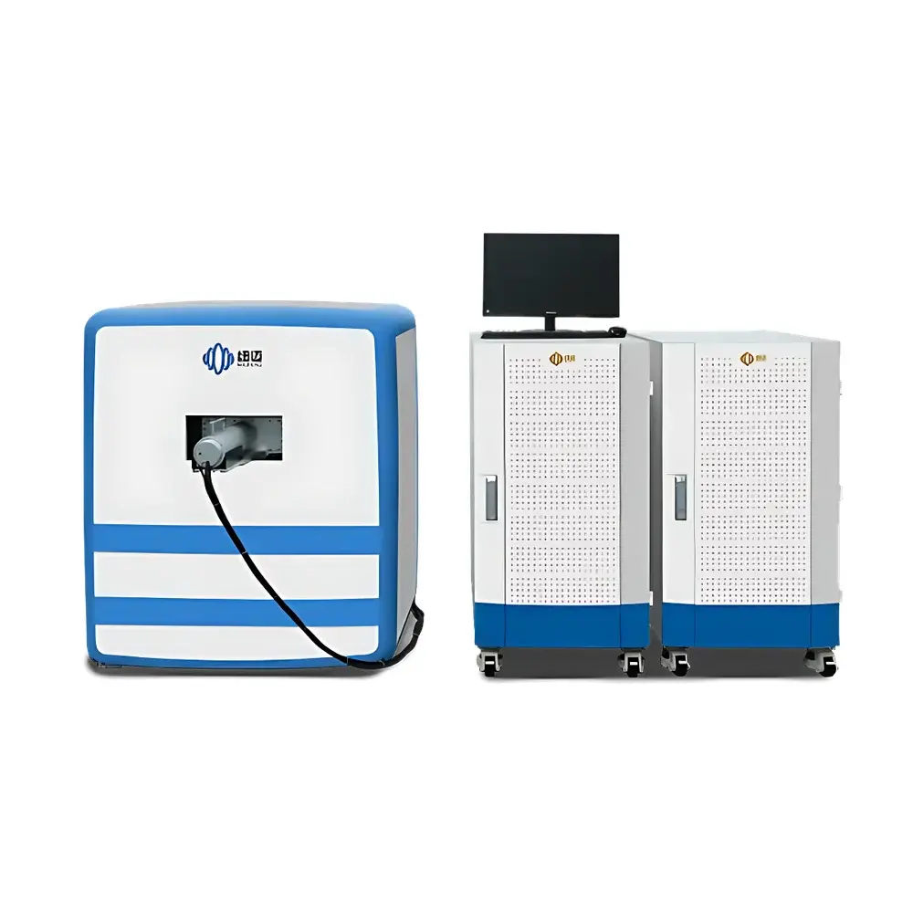
NIUMAG NM42-060H-I Small Animal MRI System
| Brand | NIUMAG |
|---|---|
| Origin | Jiangsu, China |
| Manufacturer Type | Authorized Distributor |
| Origin Category | Domestic |
| Model | NM42-060H-I |
| Instrument Type | Low-Field Nuclear Magnetic Resonance Imaging System |
| Sample Type | Solid-Liquid Dual-Mode (ex vivo tissues, live rodents: 1–350 g) |
| Magnet Type | Permanent Magnet |
| Field Strength | 1.0 T ± 0.05 T |
| Magnetic Homogeneity | ≤30 ppm over 60 mm DSV |
| Spatial Resolution | Sub-millimeter (typical in-plane: ≤150 µm, slice thickness: ≥0.5 mm) |
| Imaging Sequences | T1-weighted, T2-weighted, Proton Density-weighted, Fat/Water Separation (Dixon), Diffusion-Weighted Imaging (DWI), MR Angiography (MRA) |
Overview
The NIUMAG NM42-060H-I Small Animal MRI System is a dedicated low-field nuclear magnetic resonance imaging platform engineered for preclinical in vivo and ex vivo studies in rodent models. Operating at a stable 1.0 Tesla permanent magnet field, the system leverages robust NMR physics—based on spin excitation, phase encoding, frequency encoding, and signal reconstruction via Fourier transformation—to generate high-fidelity anatomical and functional contrast without ionizing radiation. Designed specifically for laboratory-scale translational research, it supports longitudinal monitoring of disease progression, therapeutic response, and biomaterial biodistribution in mice and rats (1–350 g). Its permanent magnet architecture eliminates cryogenic helium dependency, RF shielding room requirements, and associated infrastructure costs—making it uniquely suited for core facilities, shared imaging labs, and resource-constrained academic environments where operational simplicity and regulatory compliance are critical.
Key Features
- Permanent magnet design: Zero liquid helium consumption; no quench risk; ambient-temperature operation with passive thermal stabilization.
- High-field stability and homogeneity: 1.0 T ± 0.05 T field strength with ≤30 ppm homogeneity over a 60 mm diameter spherical volume (DSV), enabling reproducible quantitative relaxation mapping (T1, T2).
- Sub-millimeter spatial resolution: Achieves typical in-plane resolution of ≤150 µm and slice thickness down to 0.5 mm under optimized acquisition protocols—sufficient for cortical layer differentiation, tumor margin delineation, and ventricular morphology assessment.
- Multi-contrast imaging capability: Native support for T1w, T2w, PDw, fat/water separation (Dixon), diffusion-weighted imaging (DWI), and time-of-flight (TOF) MRA—enabling comprehensive tissue characterization without hardware modification.
- Integrated physiological monitoring interface: Compatible with external respiratory gating, ECG synchronization, and temperature control units for motion-compensated longitudinal studies.
Sample Compatibility & Compliance
The NM42-060H-I accommodates both live and ex vivo specimens—including intact anesthetized mice/rats, isolated organs (e.g., brain, heart, liver), tissue slices, and contrast agent formulations (e.g., Gd-based chelates, iron oxide nanoparticles). It complies with ISO/IEC 17025 general requirements for competence of testing and calibration laboratories, and its imaging protocols align with widely adopted preclinical MRI standards including the NIH/NIBIB Preclinical Imaging Guidelines and the European Federation of Organisations for Medical Physics (EFOMP) recommendations for small animal MRI QA. Data acquisition and storage adhere to DICOM 3.0 Part 10 file structure, ensuring interoperability with PACS, OsiriX, 3D Slicer, and other FDA-cleared or CE-marked analysis platforms. All sequence parameters and acquisition logs are timestamped and exportable to support GLP-compliant study documentation.
Software & Data Management
The system runs on NIUMAG’s proprietary Paravision-compatible software suite, featuring a graphical pulse sequence editor, real-time image preview, automated shimming, and batch-processing pipelines for T1/T2 mapping, ADC calculation, and ROI-based quantification. Raw k-space data is stored in vendor-neutral ISMRM Raw Data Format (ISMRMD), facilitating third-party reconstruction and machine learning model training. Audit trails record all user actions—including parameter changes, scan start/stop times, and operator ID—meeting 21 CFR Part 11 requirements for electronic records and signatures when deployed in regulated environments. Export options include NIfTI-1, MATLAB .mat, and CSV formats for statistical analysis in R, Python (NiBabel, PyTorch), or SPSS.
Applications
- Oncology: Longitudinal tracking of orthotopic glioblastoma, mammary carcinoma, and metastatic burden; evaluation of anti-angiogenic therapy efficacy via dynamic contrast-enhanced (DCE) MRI.
- Neuroscience: In vivo structural and functional assessment of stroke models, Alzheimer’s transgenic mice (e.g., APP/PS1), and neuroinflammation using T2*-weighted susceptibility mapping.
- Nanomedicine: Quantitative relaxivity (r1/r2) determination of novel contrast agents; spatiotemporal tracking of iron oxide nanoparticle accumulation in lymph nodes or tumors.
- Cardiovascular research: Cine-MRI for left ventricular ejection fraction (LVEF) and wall motion analysis in murine myocardial infarction models.
- Drug delivery: Visualization and pharmacokinetic modeling of liposomal or polymer-based theranostic carriers using dual-contrast (T1+T2) acquisition schemes.
FAQ
Is the NM42-060H-I compliant with FDA or CE regulatory pathways for preclinical imaging?
Yes—the system is classified as a Class I non-invasive diagnostic device under EU MDR Annex XVI and meets essential requirements for electromagnetic compatibility (EN 61326-1) and safety (EN 61000-6-3). While not intended for human use, its DICOM output and audit trail functionality support FDA-submitted IND/IDE packages requiring preclinical imaging evidence.
Can the system perform quantitative T1 and T2 mapping?
Yes—multi-echo spin echo (MESE) and variable repetition time (VTR) inversion recovery sequences are included, with pixel-wise fitting algorithms generating parametric maps directly in the acquisition software.
What is the maximum animal weight supported for in vivo scanning?
The system accepts live rodents up to 350 g (e.g., adult Sprague-Dawley rats) using adjustable cradles and integrated respiratory monitoring; ex vivo samples have no upper mass limit within the 60 mm DSV.
Does NIUMAG provide sequence development support for custom pulse sequences?
Yes—NIUMAG offers C++ SDK access and technical consultation for academic collaborators developing advanced sequences (e.g., compressed sensing, MR fingerprinting) under collaborative research agreements.
How is system calibration maintained over time?
Automated daily shimming routines, field probe-based drift monitoring, and quarterly QA phantom scans (using ASTM F2782-19 reference phantoms) ensure long-term geometric accuracy and intensity stability—documentation templates are provided for internal QA records.
Related Products