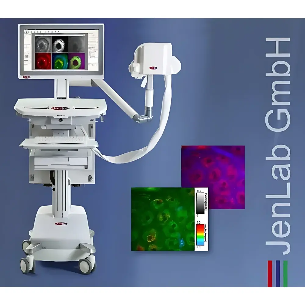
JenLab MPTcompact Compact In Vivo Multiphoton Tomography System
| Brand | JenLab |
|---|---|
| Origin | Germany |
| Manufacturer Type | Authorized Distributor |
| Origin Category | Imported |
| Model | MPTcompact |
| Pricing | Upon Request |
Overview
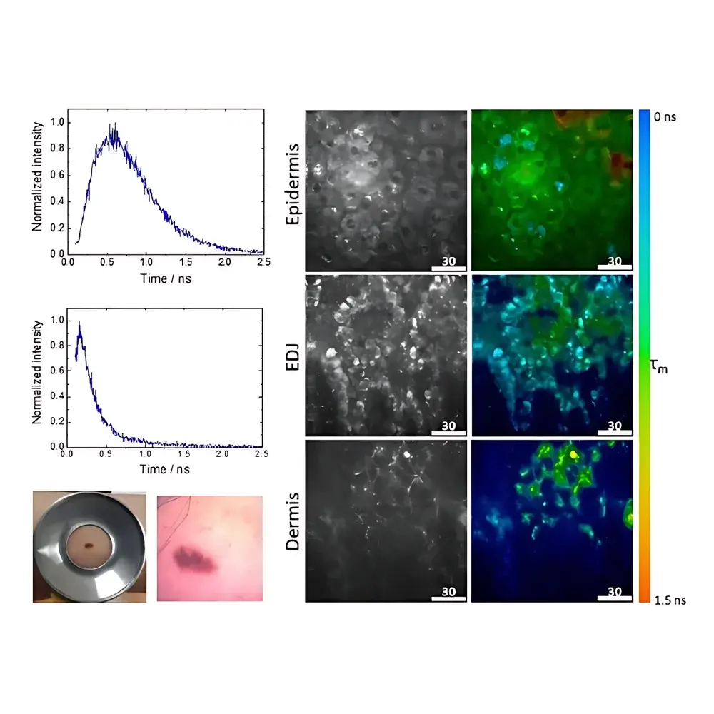
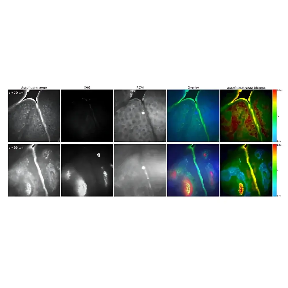
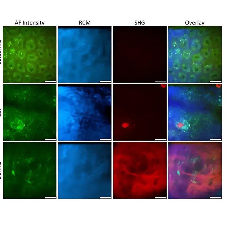
The JenLab MPTcompact is a CE-certified Class IIa medical device engineered for high-resolution, label-free, in vivo optical biopsy using multiphoton tomography (MPT). Based on near-infrared (NIR) femtosecond laser excitation at 780 nm, the system enables simultaneous detection of endogenous autofluorescence (AF) and second-harmonic generation (SHG) signals from unstained biological tissue. This dual-modal contrast mechanism leverages intrinsic molecular signatures—such as NAD(P)H, flavins, porphyrins, elastin, melanin, and fibrillar collagen—without exogenous dyes or contrast agents. The optical sectioning principle relies on nonlinear excitation confined to the focal volume, ensuring inherent optical sectioning capability, minimal photodamage, and deep-tissue penetration up to 350 µm into human skin. Designed for clinical dermatology and translational research, the MPTcompact delivers subcellular spatial resolution (<0.5 µm lateral, <2 µm axial) and picosecond temporal resolution for fluorescence lifetime imaging (FLIM), enabling quantitative assessment of cellular metabolism, extracellular matrix (ECM) architecture, and epidermal–dermal junction morphology in physiological context.
Key Features
- Integrated compact fiber-based femtosecond laser (780 ± 5 nm, pulse width <90 fs at source, 150–250 fs at sample; repetition rate 50 ± 1 MHz; average power tunable 1–50 mW at focus)
- Dual-channel detection with single-photon-sensitive photomultiplier tubes (PMTs) optimized for simultaneous AF and SHG acquisition
- High-NA oil-immersion objective (40×, NA 1.3) with 200 µm working distance and diffraction-limited focusing
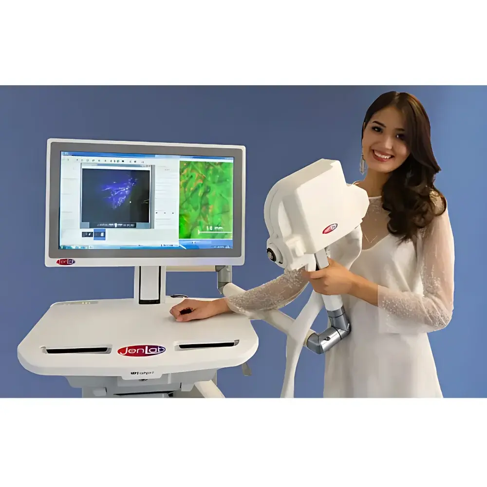
- 360° articulating measurement head mounted on precision robotic arm, enabling ergonomic positioning and repeatable probe alignment
- Multi-modal imaging suite: AF imaging, SHG imaging, FLIM/OMI (Optical Metabolic Imaging), confocal reflectance laser scanning microscopy (RLSM), and digital dermoscopy
- Full-field white-light imaging for anatomical context registration and lesion mapping
- Compact footprint (750 × 900 × 1600 mm) and modular design suitable for clinical rooms, GMP-compliant labs, and mobile research units
Sample Compatibility & Compliance
The MPTcompact is validated for non-invasive, real-time imaging of human skin *in vivo*, including epidermis, dermo-epidermal junction (DEJ), and upper reticular dermis. It supports longitudinal monitoring of melanocytic lesions, inflammatory responses, wound healing dynamics, and ECM remodeling. As a Class IIa medical device under EU MDR 2017/745, it complies with essential requirements for safety, performance, electromagnetic compatibility (EMC), and laser safety per DIN EN 60825-1 (Laser Class 1M). The system meets ISO 13485 quality management standards for medical device manufacturing and distribution. Data acquisition workflows support audit trails and electronic signature compliance per FDA 21 CFR Part 11 when integrated with validated LIMS or clinical trial software environments. All imaging protocols adhere to ISO/IEC 17025 principles for method validation in diagnostic applications.
Software & Data Management
Acquisition and analysis are performed via JenLab’s proprietary MPT software platform, which provides real-time visualization, automated image stitching, spectral unmixing (for multi-fluorophore separation), and FLIM decay curve fitting using multi-exponential models. Quantitative metrics—including NAD(P)H/FAD redox ratio, collagen/elastin SHG-to-AF intensity ratio (CEAR), fluorescence lifetime τm, and spatial heterogeneity indices—are exportable in CSV, TIFF, and OME-TIFF formats. The software supports DICOM-SR export for integration into PACS systems and includes built-in tools for GLP/GMP-aligned documentation: user access control, versioned protocol templates, timestamped metadata embedding, and raw-data integrity verification. Raw time-resolved photon arrival data is stored in HDF5 format to preserve full temporal and spatial fidelity for retrospective reanalysis.
Applications
- Early detection and differential diagnosis of melanoma and non-melanoma skin cancers via morphological and metabolic profiling of melanocytes, keratinocytes, and dendritic cells
- Quantitative assessment of skin aging through elastin/collagen network degradation, epidermal thickness mapping, and mitochondrial metabolic activity (via NAD(P)H τ1/τ2 ratios)
- In vivo evaluation of topical drug penetration kinetics, sunscreen nanoparticle biodistribution, and transdermal delivery efficiency
- Longitudinal monitoring of wound healing, scar formation, and fibrotic remodeling in clinical trials and preclinical models
- Non-destructive quality control in tissue-engineered skin constructs, including 3D ECM organization and cell viability mapping
- Regulatory-compliant safety and efficacy testing of cosmeceuticals under ISO 10993-10 and OECD TG 439 guidelines
FAQ
Is the MPTcompact approved for clinical use in the EU?
Yes—the system carries CE marking as a Class IIa medical device under Regulation (EU) 2017/745 and is cleared for diagnostic imaging of skin pathologies in clinical dermatology settings.
Can the system be used for longitudinal patient monitoring?
Yes—its non-invasive, label-free operation enables repeated measurements over days, weeks, or months without tissue damage or signal quenching, supporting robust intra-patient comparative analysis.
What regulatory standards does the software meet for clinical data handling?
The acquisition software supports ALCOA+ data integrity principles and can be configured for 21 CFR Part 11 compliance when deployed in validated environments with appropriate IT infrastructure and procedural controls.
Does the system require specialized infrastructure or shielding?
No—operation requires only standard 220–230 V/50 Hz power, ambient temperature (17–25 °C), and relative humidity (20–65%). No laser interlock room or RF-shielded enclosure is needed due to its Class 1M laser classification.
How is image quantification standardized across operators and sites?
Standardized calibration routines (laser power, PMT gain, detector offset) are embedded in each protocol; all quantitative outputs include traceable metadata (e.g., excitation power at focus, objective temperature, ambient humidity) to enable cross-site reproducibility per ISO/IEC 17025 Annex A.3.


