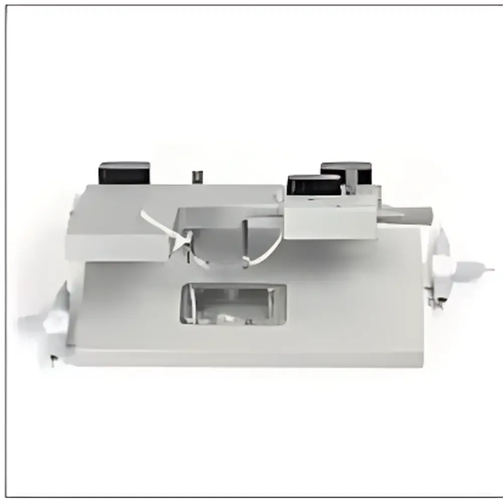
Laser Scanning Confocal Ex Vivo Microvessel Pressure-Diameter Measurement System 120CP
| Origin | USA |
|---|---|
| Manufacturer Type | Authorized Distributor |
| Origin Category | Imported |
| Model | 120CP |
| Pricing | Upon Request |
Overview
The Laser Scanning Confocal Ex Vivo Microvessel Pressure-Diameter Measurement System 120CP is a precision-engineered platform designed for quantitative biomechanical and functional assessment of isolated microvessels under near-physiological conditions. Built to integrate seamlessly with commercial laser scanning confocal microscopes (LSCM) and high-resolution upright or inverted imaging systems, the 120CP enables simultaneous real-time measurement of intraluminal pressure, vessel diameter, and fluorescent signal dynamics in pressurized arterioles and small resistance arteries (diameter >60 µm). Its operational principle relies on servo-controlled pressure regulation coupled with high-magnification optical diameter tracking—typically via edge-detection algorithms applied to confocal reflectance or fluorescence images—allowing precise quantification of myogenic tone, flow-mediated dilation, vasoconstrictor responsiveness, and endothelial-dependent relaxation. The system maintains physiological temperature (37 °C), pH (7.4), and oxygenation (95% O₂/5% CO₂) within the superfusion bath, supporting long-term viability (>4–6 h) of freshly isolated vessels from rodent, porcine, or human tissue sources.
Key Features
- Integrated pressure-clamp architecture with digital servo control for stable intraluminal pressure ranging from 0 to 150 mmHg (adjustable in 0.1 mmHg increments)
- Optimized optical interface: accommodates objective lenses with working distances as short as 100 µm, enabling high-NA immersion objectives for subcellular resolution imaging
- Dedicated low-autofluorescence, temperature-regulated superfusion chamber with dual-inlet perfusion ports and integrated gas exchange manifold
- Modular design allows direct mechanical coupling to LSCM stages without optical path obstruction or vibration transmission
- Real-time diameter feedback loop synchronized with pressure modulation and fluorescence acquisition (e.g., Ca²⁺, NO, ROS indicators)
- Compliance with ISO 13485–aligned manufacturing traceability and CE marking for in vitro diagnostic research use
Sample Compatibility & Compliance
The 120CP supports ex vivo studies of pressurized microvessels including cerebral, mesenteric, coronary, and renal resistance arteries (60–300 µm outer diameter). Vessels are cannulated at both ends using glass micropipettes and secured with nylon sutures; viability is confirmed via agonist-induced constriction and endothelium-dependent vasodilation. The system conforms to ASTM F2921–22 standards for vascular tissue preparation and testing protocols. All wetted components are autoclavable or ethylene oxide–sterilizable. Data acquisition workflows comply with GLP-aligned documentation requirements, and optional audit trail modules support FDA 21 CFR Part 11–compliant electronic records when paired with validated third-party acquisition software.
Software & Data Management
Control and analysis are performed using vendor-provided Windows-based software featuring synchronized multi-channel acquisition (pressure, diameter, fluorescence intensity, time-stamped event markers). Raw data export is supported in HDF5 and CSV formats for downstream processing in MATLAB, Python (NumPy/Pandas), or Igor Pro. The software includes built-in routines for calculating wall stress, circumferential strain, distensibility, and myogenic index (ΔD/D₀ vs. ΔP), with batch-processing capability for comparative pharmacological profiling. Optional API integration enables remote scripting and automated protocol execution in multi-user core facility environments.
Applications
- Myogenic autoregulation and pressure-induced vasoconstriction in hypertension and diabetes models
- Endothelial nitric oxide synthase (eNOS) activity and redox-sensitive signaling using DAF-FM or DHE probes
- Calcium handling in vascular smooth muscle cells via Fluo-4 or Fura-2 ratiometric imaging
- Pharmacological characterization of GPCR ligands, ion channel modulators (e.g., TRPV4, BKCa), and Rho-kinase inhibitors
- Microvascular dysfunction in aging, sepsis, and chronic kidney disease using human resistance artery biopsies
- Validation of computational hemodynamic models requiring experimentally derived passive/active mechanical parameters
FAQ
What vessel size range is compatible with the 120CP system?
Vessels with outer diameters between 60 µm and 300 µm are routinely studied; optimal performance is achieved for segments 800–1500 µm in length.
Can the system be used with two-photon microscopy?
Yes—the optical layout preserves full access to the specimen plane, supporting integration with multiphoton excitation paths and non-descanned detection configurations.
Is temperature and pH control independently adjustable?
Superfusion solution temperature is regulated via inline heater/cooler (±0.1 °C stability); pH is maintained by continuous carbogen equilibration and verified with inline micro-pH electrodes.
Does the system support simultaneous intracellular pressure and membrane potential recording?
While not natively equipped with electrophysiology hardware, the 120CP’s open I/O architecture permits synchronization with patch-clamp amplifiers and digitizers via TTL triggers and analog voltage inputs.
What validation documentation is provided for regulatory submissions?
A comprehensive Device Master Record (DMR)-aligned technical file—including calibration certificates, risk analysis (ISO 14971), and software verification reports—is available upon request for preclinical study audits.


