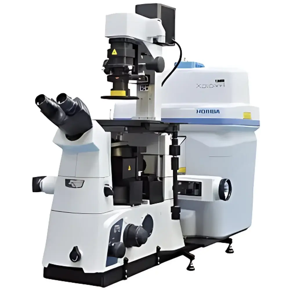
HORIBA LabRAM XploRA INV Inverted Confocal Raman Microscope
| Brand | HORIBA |
|---|---|
| Origin | France |
| Model | LabRAM XploRA INV |
| Instrument Type | Inverted Confocal Raman Microscope |
| Laser Options | 532 nm, 638 nm, 785 nm (3 internal lasers) |
| Grating Options | 600, 1200, 1800, 2400 gr/mm (motorized auto-switching) |
| Spectral Range | 250–10,000 cm⁻¹ (configurable per laser/grating combination) |
| Laser Power Control | Multi-step neutral density attenuation |
| Automation | Auto-focus, auto-exposure, auto-calibration, self-diagnostic routine |
| Software Compliance | FDA 21 CFR Part 11-ready audit trail, GLP/GMP-compatible workflow logging |
| Optional Modules | DuoScan™ high-speed Raman imaging, SWIFT™ fast spectral acquisition, epifluorescence integration, polarization optics, precision XYZ stage, environmental stages (cryo/heated/biological incubation), AFM/Raman coupling interface, fiber-optic probe compatibility |
Overview
The HORIBA LabRAM XploRA INV is an inverted confocal Raman microscope engineered for high-fidelity molecular characterization of delicate, hydrated, or live biological specimens under native or near-physiological conditions. Unlike conventional upright configurations, its inverted optical architecture places the objective lens beneath the sample—enabling unobstructed access to the top surface for micromanipulation, fluidic interfacing, or integration with auxiliary instrumentation such as optical tweezers, patch-clamp rigs, or microinjection systems. The system operates on the principle of inelastic light scattering (Raman effect), where monochromatic laser excitation induces vibrational mode shifts in molecular bonds, yielding fingerprint-like spectral signatures with sub-micron spatial resolution in confocal mode. Its modular design supports both steady-state point spectroscopy and hyperspectral Raman imaging across a broad wavenumber range (250–10,000 cm⁻¹), with selectable excitation wavelengths (532, 638, 785 nm) optimized for minimizing fluorescence background and photodamage in biological matrices.
Key Features
- Research-grade inverted microscope platform with long-working-distance objectives and parfocal alignment for seamless transition between brightfield, phase contrast, and Raman imaging.
- Triple-laser engine with motorized wavelength selection—enables rapid switching between 532 nm (high sensitivity), 638 nm (balanced signal-to-noise), and 785 nm (reduced autofluorescence in tissues/cells).
- Four-grating turret (600, 1200, 1800, 2400 gr/mm) with automated alignment—provides user-selectable spectral resolution (down to ~1 cm⁻¹ FWHM at 532 nm) and dispersion trade-offs without manual realignment.
- Fully automated operational sequence: real-time auto-focus via motorized Z-stage feedback, exposure time optimization based on signal saturation thresholds, built-in NIST-traceable calibration using silicon reference peak (520.7 cm⁻¹), and daily self-diagnostic protocol verifying laser stability, grating positioning, and detector linearity.
- DuoScan™ technology: galvanometric mirror-based scanning enabling sub-second pixel dwell times for large-area Raman mapping (e.g., 100 × 100 µm² at 1 µm step size in <5 min), compatible with correlative Raman-fluorescence-PL imaging.
- Open mechanical architecture accommodating third-party accessories—including motorized XYZ stages with sub-100 nm repeatability, temperature-controlled stages (−190 °C to +600 °C), humidity chambers, and AFM-Raman hybrid probes with synchronized topography-spectra acquisition.
Sample Compatibility & Compliance
The LabRAM XploRA INV accommodates diverse sample formats: live cells in culture dishes (glass-bottom or polymer substrates), tissue sections on standard microscope slides, 3D organoids in Matrigel, bacterial biofilms on functionalized surfaces, and microfluidic devices with integrated optical windows. Its inverted geometry eliminates interference from coverslip thickness variations and permits direct immersion objectives for water- or oil-immersion measurements. All hardware and software modules comply with ISO/IEC 17025 requirements for analytical instrument validation. The embedded QC routines satisfy ASTM E1840-22 (Standard Practice for Calibration of Raman Spectrometers) and support GLP/GMP documentation workflows, including electronic signatures, version-controlled method files, and 21 CFR Part 11-compliant audit trails for raw data, processing parameters, and operator actions.
Software & Data Management
Control and analysis are executed via HORIBA’s LabSpec 6 software—a modular suite supporting spectral preprocessing (cosmic ray removal, baseline correction, vector normalization), multivariate analysis (PCA, cluster analysis, MCR-ALS), and quantitative modeling (peak fitting, intensity ratio mapping). Raw spectra are stored in HDF5 format with embedded metadata (laser power, grating ID, exposure time, objective magnification, stage coordinates). Batch processing pipelines enable automated quality control of multi-sample datasets, while API access (Python/C++) allows integration into LIMS or custom data lakes. The GO! assistant provides context-aware guidance for method setup, reducing training time for new users without compromising analytical rigor.
Applications
- Subcellular chemical mapping of lipid droplets, protein aggregates, or drug distribution in fixed and live mammalian cells.
- Label-free identification of bacterial strains and antibiotic resistance markers via spectral biomarkers (e.g., nucleic acid vs. peptidoglycan band ratios).
- In situ monitoring of extracellular matrix remodeling during osteogenesis or chondrogenesis in 3D scaffolds.
- Correlative analysis combining Raman spectral fingerprints with epifluorescence localization of tagged proteins or organelles.
- Process analytical technology (PAT) applications: real-time reaction monitoring in microreactors or crystallization studies under controlled thermal/humid conditions.
- Forensic analysis of trace evidence (e.g., pigment identification in single textile fibers or ink layers in questioned documents).
FAQ
Is the LabRAM XploRA INV compatible with live-cell imaging under CO₂-controlled environments?
Yes—the open frame design permits integration with commercial incubation chambers and gas controllers; optional heated stage with humidity regulation maintains physiological conditions during extended acquisitions.
Can spectral data be exported for third-party chemometric analysis?
Yes—LabSpec 6 exports spectra in ASCII, CSV, and standardized JCAMP-DX formats; HDF5 exports include full metadata for reproducible reprocessing.
Does the system support regulatory submissions under FDA or EMA guidelines?
Yes—audit trail configuration, electronic signature support, and method validation templates align with ICH M10 and FDA Bioanalytical Method Validation Guidance requirements.
What is the typical spatial resolution achievable in confocal mode?
Lateral resolution is diffraction-limited (~0.5 µm at 532 nm with 100× oil objective); axial resolution is ~1–2 µm depending on pinhole size and refractive index matching.
How is laser-induced sample damage mitigated during long-duration mapping?
Multi-level ND filters, real-time power monitoring, and adaptive exposure algorithms dynamically adjust irradiance to maintain sub-threshold phototoxicity levels—validated via cell viability assays (e.g., Calcein AM/PI staining).