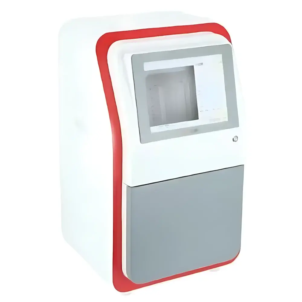
Jiapeng JP-K3900Plus Chemiluminescence Gel Imaging System
| Brand | Jiapeng |
|---|---|
| Origin | Shanghai, China |
| Model | JP-K3900Plus |
| Imaging Type | Multicolor Fluorescence & Chemiluminescence Gel Imager |
| CCD Resolution | 9 MP (3000 × 3000) |
| Bit Depth | 16-bit |
| Dynamic Range | 0–4.8 OD |
| CCD Sensor Size | 4/3" (SONY IMX533CLK-D) |
| Pixel Size | 5.4 µm × 5.4 µm |
| Cooling Temperature | −45 °C (ΔT ≈ −65 °C vs. ambient) |
| Quantum Efficiency | >75% |
| Lens | F/0.95 Fixed Focus (optional F/0.8) |
| Optical Zoom | 8–48 mm (6×) |
| Detection Sensitivity | ≥0.02 ng Protein (Western blot), ≥0.05 ng DNA (ethidium bromide-stained) |
| Signal-to-Noise Ratio | ≥56 dB |
| Transmission UV Sources | 300 nm (standard), 254/365 nm (optional reflectance) |
| UV Transilluminator Area | 200 × 200 mm |
| White Light Transmission Panel | Uniform LED-based, integrated |
| Filter Wheel | Motorized 5-position (optional 10-position), with standard bandpass filters at 535, 590, 605, and 699 nm |
| Imaging Area Options | 9.5 × 9.5 cm and 12.5 × 12.5 cm (dual-stage drawer platform) |
| Power Supply | AC 220 V, 50/60 Hz |
| Dimensions (W × D × H) | 390 × 440 × 735 mm |
| Weight | 18.3 kg |
Overview
The Jiapeng JP-K3900Plus Chemiluminescence Gel Imaging System is a high-performance, fully automated platform engineered for quantitative detection and analysis of nucleic acids, proteins, and reporter-labeled biomolecules across multiple optical modalities. It integrates cooled scientific-grade CMOS imaging, precision optical filtration, and programmable illumination control to support chemiluminescence (CL), bioluminescence (BL), ultraviolet (UV) transillumination, multi-channel fluorescence (blue, green, red, near-infrared), and white-light transmission/reflection imaging—all within a single darkroom architecture. The system employs a back-illuminated SONY IMX533CLK-D sensor (4/3″ format, 9 MP resolution) with thermoelectric cooling to −45 °C—enabling low-dark-current acquisition (75%) essential for detecting weak CL signals from Western blots or low-abundance luciferase assays. Its 16-bit digitization (0–65,535 intensity levels) and calibrated optical density range of 0–4.8 OD ensure linear quantification across four orders of magnitude, meeting requirements for publication-grade densitometry in peer-reviewed life science research.
Key Features
- Cooled scientific imaging sensor with deep thermoelectric cooling (−45 °C nominal, ΔT ≈ −65 °C below ambient) and ultra-low dark current for extended exposure without thermal noise accumulation
- F/0.95 high-transmission fixed-focus lens optimized for maximum photon capture in low-light chemiluminescent applications; optional F/0.8 variant available for enhanced sensitivity
- Dual-stage drawer-style sample platform supporting two discrete imaging areas (9.5 × 9.5 cm and 12.5 × 12.5 cm), enabling rapid switching between small-format membranes and full-size gels without realignment
- Motorized 5-position filter wheel (upgradeable to 10-position) with precise spectral control; standard interference filters centered at 535 nm (green fluorescence), 590 nm (orange), 605 nm (red), and 699 nm (far-red/NIR)
- Integrated uniform white-light transmission panel and top-mounted high-stability white LED source for Coomassie- and Ponceau-stained gel documentation
- Programmable UV transilluminator (300 nm standard; optional 254/365 nm reflectance modules) with 200 × 200 mm active area and safety interlock circuitry
- Onboard 12-inch industrial LCD touchscreen interface with embedded acquisition software; also supports remote operation via Ethernet-connected Windows workstation
Sample Compatibility & Compliance
The JP-K3900Plus accommodates standard electrophoretic formats including mini- and midi-gels (up to 15 × 15 cm), nitrocellulose and PVDF membranes (up to 12 × 12 cm), microplates (6–96-well), and tissue sections mounted on glass slides. Its modular illumination design ensures compatibility with common detection chemistries—including HRP- and AP-conjugated secondary antibodies, luminol/peroxide-based substrates (e.g., ECL, SuperSignal), luciferin/luciferase systems, SYBR Safe, Cy dyes, Alexa Fluor conjugates, and ethidium bromide. The system supports GLP-compliant workflows through audit-trail-enabled acquisition logs, user-access controls, and timestamped metadata embedding (EXIF-compatible). While not pre-certified to FDA 21 CFR Part 11, its software architecture allows configuration for electronic signature and data integrity requirements when deployed in regulated QC environments.
Software & Data Management
Acquisition and analysis are performed using Jiapeng’s proprietary GelCapture Pro v5.x software, compatible with Windows 10/11 (64-bit). The application provides real-time preview, auto-exposure optimization, background subtraction, lane/band detection with molecular weight calibration, and multiplex ratio analysis across fluorescence channels. All images are saved in lossless TIFF format with embedded metadata (exposure time, lens aperture, filter ID, temperature, OD calibration coefficients). Batch processing supports ROI-based quantification, normalization to loading controls, and export to CSV, Excel, or PDF reports. Raw image archives can be synchronized to network storage or LIMS via configurable SMB/CIFS protocols. Software updates are delivered via secure HTTPS channel with SHA-256 signature verification.
Applications
- Quantitative Western blot analysis using chemiluminescent substrates (e.g., detection of phosphorylated kinases at sub-nanogram levels)
- Fluorescent multiplexing of protein expression profiles using far-red and NIR dyes to minimize autofluorescence interference
- DNA fragment sizing and purity assessment in agarose and polyacrylamide gels stained with SYBR Gold or GelRed
- Reporter gene assays (luciferase, GFP variants) in cell lysates and live-cell imaging plates
- Colony hybridization and Southern/Northern blot documentation under UV and visible light
- Stain-free total protein normalization using internal reference standards imaged under 300 nm UV
FAQ
What is the minimum detectable amount of protein using chemiluminescence on this system?
The JP-K3900Plus achieves ≥0.02 ng sensitivity for HRP-conjugated antibodies on nitrocellulose membranes under standard ECL conditions with 1–5 minute exposures.
Can the system perform simultaneous dual-color fluorescence imaging?
No—it acquires multichannel fluorescence sequentially using the motorized filter wheel; however, spectral unmixing and co-localization analysis are supported in post-processing.
Is the UV transilluminator suitable for DNA visualization without gel extraction?
Yes—the 300 nm transilluminator provides optimal excitation for ethidium bromide and SYBR-class dyes while minimizing DNA damage compared to 254 nm sources.
Does the system support automated focus calibration?
Yes—autofocus routines are implemented via contrast-based edge detection across the sample plane, with manual fine-tuning available via on-screen slider.
What file formats are supported for data export and integration with third-party analysis tools?
TIFF (with metadata), PNG, JPEG, and CSV for intensity tables; export modules for ImageJ/Fiji, GraphPad Prism, and Proteome Discoverer are documented in the SDK.


