Jiapeng JP-K600 Chemiluminescence Gel Imaging System
| Brand | Jiapeng |
|---|---|
| Origin | Shanghai, China |
| Model | JP-K600 |
| Instrument Type | Chemiluminescence Gel Imaging System |
| CCD Resolution | 9 MP (3000 × 3000) |
| Bit Depth | 16-bit |
| Dynamic Range | >4.0 OD (4.8 OD typical) |
| CCD Sensor Size | 4/3" (SONY IMX533CLK-D) |
| Pixel Size | 5.4 µm × 5.4 µm |
| Cooling Temperature | −45 °C (ΔT ≈ −65 °C vs ambient) |
| Lens Aperture | f/0.95 fixed-focus macro lens |
| Detection Sensitivity | <20 pg double-stranded DNA stained with ethidium bromide |
| Signal-to-Noise Ratio | ≥56 dB |
| White Light Transmission Area | 200 × 200 mm |
| UV Transillumination | 300 nm (transmission), optional 254/365 nm (reflection) |
| Filter Wheel | 5-position motorized (optional 10-position) |
| Standard Emission Filter | 590 nm |
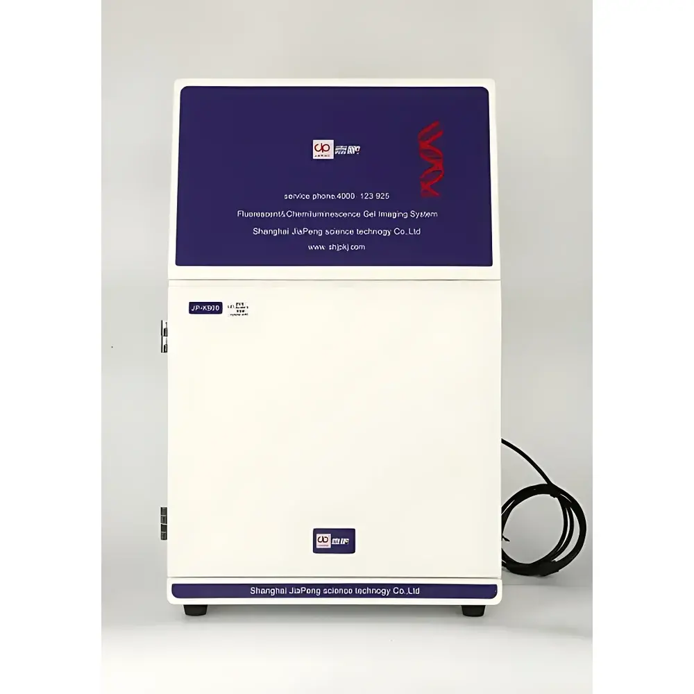
| Dimensions (W×D×H) | 390 × 440 × 735 mm |
| Weight | 18.3 kg |
Overview
The Jiapeng JP-K600 Chemiluminescence Gel Imaging System is a fully integrated, high-sensitivity digital imaging platform engineered for quantitative detection and analysis of nucleic acids and proteins across multiple modalities—including UV transilluminated gel documentation, chemiluminescent Western blot imaging, bioluminescent reporter assays, and fluorescence-based membrane detection. At its core lies a thermoelectrically cooled scientific-grade CMOS sensor (SONY IMX533CLK-D), optimized for low-noise, high-quantum-efficiency photon capture under ultra-low-light conditions. Unlike conventional CCD-based systems, this back-illuminated 4/3-inch sensor delivers exceptional quantum efficiency (>75%) and deep cooling (−45 °C stabilized, up to 65 °C below ambient), enabling reliable integration of weak, transient chemiluminescent signals—critical for detecting low-abundance targets in immunoblotting or luciferase-based assays. The system operates on a rigid, light-tight enclosure architecture with precisely calibrated optical path geometry, ensuring spatial fidelity, repeatability, and minimal stray-light interference during long-exposure acquisitions.
Key Features
- Thermoelectrically cooled 9 MP (3000 × 3000) scientific CMOS sensor with 16-bit digitization and >4.0 OD dynamic range (up to 4.8 OD), supporting pixel binning (1×1 to 8×8) for adaptive signal-to-noise optimization.
- f/0.95 high-transmission, fixed-focus macro lens with diffraction-limited resolution—designed specifically for uniform field illumination and minimal vignetting across the full imaging area.
- Dual-format sample stage: standard drawer-style dual-position platform with manual height adjustment; optional motorized Z-axis stage for automated focus calibration and multi-layer stack acquisition.
- Integrated white-light transmission illuminator (200 × 200 mm) with uniformity >95%, compliant with ISO 15739:2013 for flat-field consistency in densitometric quantification.
- Motorized 5-position filter wheel (standard), programmable via software interface; supports spectral selection for chemiluminescence (e.g., 590 nm bandpass), fluorescence (e.g., FITC, Cy3, Cy5), and UV reflectance applications.
- User-configurable auto-shutdown timer (0–60 min) for excitation sources; dark-current suppression (<0.0005 e⁻/pixel/s) validated per EMVA 1288 standards.
Sample Compatibility & Compliance
The JP-K600 accommodates standard electrophoretic formats including mini- and midi-gels (up to 12.5 × 12.5 cm), PVDF and nitrocellulose membranes, microplates (6–96-well), and tissue sections mounted on glass slides. Its optical design conforms to IEC 61000-6-3 (EMC emissions) and IEC 61010-1 (safety requirements for laboratory equipment). For regulated environments, image acquisition metadata—including exposure time, gain, lens aperture, filter position, temperature log, and user ID—is embedded in TIFF headers and exportable in FAIR-compliant formats. Audit trails and user-access controls align with GLP and GMP documentation expectations; raw image files retain unprocessed 16-bit linear intensity values required for retrospective reanalysis per FDA 21 CFR Part 11 guidelines.
Software & Data Management
The bundled ImageLab Pro software provides end-to-end workflow support—from real-time preview and auto-exposure calculation to background subtraction, lane/band identification, molecular weight estimation, and relative quantification using internal loading controls. All processing steps are scriptable and reproducible; batch operations support multi-condition comparisons across replicate blots or time-series experiments. Export options include annotated TIFF, PNG, PDF, and CSV (for intensity tables); DICOM-SR compatibility is available upon request for institutional PACS integration. Data integrity safeguards include SHA-256 checksum generation, timestamped version history, and optional network storage synchronization with NAS or LIMS endpoints.
Applications
- Quantitative Western blot analysis using HRP- or AP-conjugated secondary antibodies with enhanced chemiluminescent substrates (e.g., Luminata Crescendo, ECL Prime).
- DNA/RNA gel documentation under UV (300 nm transillumination) and visible light, including ethidium bromide-, SYBR Safe-, and GelRed-stained samples.
- Bioluminescent imaging of luciferase-expressing cell lines or animal tissues (e.g., firefly, Renilla, NanoLuc reporters) without external excitation.
- Fluorescent membrane imaging with near-infrared dyes (e.g., IRDye 680RD, Alexa Fluor 647) when equipped with appropriate excitation/emission filters.
- Densitometric validation of qPCR product integrity, CRISPR editing efficiency (T7E1 assay), and RNA integrity number (RIN) estimation from denaturing gels.
FAQ
What is the minimum detectable amount of DNA using ethidium bromide staining?
The JP-K600 achieves sub-20 pg detection sensitivity for double-stranded DNA visualized with ethidium bromide under standard UV transillumination (300 nm), verified using Lambda-HindIII ladder dilution series per ISO 10993-12 protocols.
Can the system perform multiplex fluorescent imaging?
Yes—when configured with optional excitation sources (e.g., 470 nm LED, 530 nm LED) and corresponding emission filters, the JP-K600 supports two-color fluorescence detection on membranes, provided spectral separation exceeds 30 nm FWHM.
Is remote operation supported?
The system supports LAN-based control via TCP/IP; full GUI mirroring and command-line acquisition scripting (Python API) are included in the Enterprise license tier.
How is focus calibrated across different sample heights?
The standard drawer platform includes mechanical stops for 2 predefined positions (gel vs. membrane); the optional motorized Z-stage enables software-driven focus mapping and autofocus routines based on contrast gradient maximization.
Does the software comply with 21 CFR Part 11 requirements?
ImageLab Pro offers optional 21 CFR Part 11 add-on module featuring electronic signatures, role-based access control, immutable audit logs, and electronic record retention policies—validated per IQ/OQ protocols upon installation.


