Thermo Fisher Axia ChemiSEM Intelligent Tungsten-Filament Scanning Electron Microscope
| Brand | Thermo Fisher |
|---|---|
| Origin | Netherlands |
| Model | Axia ChemiSEM |
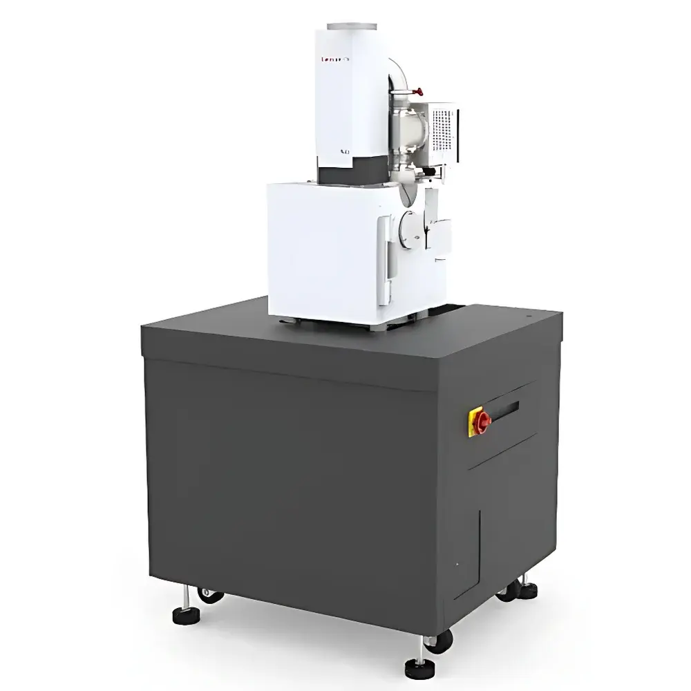
| Instrument Type | Floor-standing Conventional SEM |
| Electron Source | Pre-aligned Tungsten Filament |
| Secondary Electron (SE) Resolution | 3.0 nm @ 30 kV (HV), 8.0 nm @ 3 kV (HV) |
| Backscattered Electron (BSE) Resolution | 7.0 nm @ 3 kV (HV, decelerated mode) |
| Magnification Range | 5× to 1,000,000× |
| Accelerating Voltage | 200 V – 30 kV |
| Probe Current | Up to 2 µA, continuously adjustable |
| Low Vacuum Mode | Up to 150 Pa |
| Working Distance for EDS | 10 mm |
| EDS Take-off Angle | 35° |
| Chamber Internal Width | 280 mm |
| Number of Ports | 10 |
| Stage | 5-axis motorized (X=120 mm, Y=120 mm, Z=55 mm, Tilt=–15° to +90°, Rotation=360° continuous) |
| Max Sample Dimensions | 240 × 280 × 128 mm³ |
| Max Sample Weight | 10 kg |
| Detectors | In-chamber IR navigation camera, Nav-Cam+™ color optical camera, ETD (Everhart-Thornley SE detector), retractable BSED, TrueSight X EDS with ChemiSEM technology |
| Control System | Windows 10 OS, 24″ LCD display (1920×1200), customizable GUI supporting quad-image real-time display, full Undo/Redo functionality |
Overview
The Thermo Fisher Axia ChemiSEM is a floor-standing, intelligent tungsten-filament scanning electron microscope engineered for routine and advanced materials characterization in industrial quality control, academic research, and failure analysis laboratories. Unlike conventional SEMs requiring extensive operator expertise for alignment and optimization, the Axia ChemiSEM integrates a pre-aligned tungsten electron source with a robust, maintenance-optimized column architecture—enabling rapid startup, stable beam performance, and consistent imaging across variable operating conditions. Its core innovation lies in ChemiSEM technology: a hardware-software co-engineered platform that synchronizes real-time energy-dispersive X-ray spectroscopy (EDS) acquisition with primary electron scanning. This eliminates sequential acquisition workflows, delivering spatially registered morphological and compositional data in a single pass—without compromising resolution or signal fidelity. Designed for high reproducibility under GLP-compliant workflows, the system operates across high vacuum (HV), low vacuum (LV), and beam deceleration modes, making it uniquely suited for insulating, hydrated, or beam-sensitive specimens without mandatory conductive coating.
Key Features
- Pre-aligned tungsten filament source: Delivers long-term emission stability and reduced filament replacement frequency; no routine gun alignment required.
- ChemiSEM real-time EDS integration: Simultaneous acquisition of secondary electron (SE), backscattered electron (BSE), and characteristic X-ray signals during raster scanning—enabling immediate elemental mapping alongside topographic imaging.
- Full-access chamber design: Wide-opening front door (280 mm internal width) and motorized 5-axis stage support large, heavy samples (up to 10 kg, 240 × 280 × 128 mm³) with precise positioning and tilt/rotation for cross-sectional and angular analysis.
- Dual-mode imaging capability: High-vacuum operation (down to 3.0 nm SE resolution at 30 kV) and low-vacuum mode (up to 150 Pa) for charge mitigation on non-conductive samples; beam deceleration mode enhances surface sensitivity and reduces penetration depth at low kV.
- Nav-Cam+™ optical navigation system: Integrated color CCD camera provides real-time optical preview and coordinate-based sample targeting—reducing time-to-region-of-interest by >40% versus traditional manual stage movement.
- TrueSight X EDS detector: Silicon drift detector (SDD) with 35° take-off angle and 10 mm working distance optimized for high count-rate, low-noise spectral collection; supports quantitative standardless and standards-based analysis per ISO 16574 and ASTM E1508.
Sample Compatibility & Compliance
The Axia ChemiSEM accommodates a broad spectrum of solid-state materials—including metals, ceramics, polymers, geological specimens, biological tissues (fixed or critical-point dried), and electronic components—without mandatory sputter coating in most cases. Its low-vacuum capability (up to 150 Pa) permits direct imaging of moderately outgassing or mildly hydrated samples, while the beam deceleration mode mitigates charging on thin films and nanocomposites. The system meets mechanical and electrical safety requirements per IEC 61010-1 and electromagnetic compatibility per EN 61326-1. Software audit trails, user access controls, and electronic signature support align with FDA 21 CFR Part 11 for regulated environments. Routine operation complies with ISO/IEC 17025 documentation standards for testing laboratories.
Software & Data Management
Controlled via a Windows 10-based interface with a fully customizable GUI, the Axia ChemiSEM software enables concurrent display of up to four synchronized image streams (SE, BSE, EDS map, optical preview). All acquisition parameters—including kV, probe current, dwell time, and EDS live-time—are logged automatically with timestamped metadata. The integrated Undo/Redo engine allows iterative parameter refinement without re-scanning. Quantitative EDS analysis adheres to ZAF and φ(ρz) matrix correction models; spectral libraries conform to IUPAC-recommended peak assignments. Raw data (image stacks, spectra, maps) are stored in vendor-neutral formats (TIFF, .emsa, .msa) for third-party processing. Optional Thermo Scientific Velox software extends capabilities to automated particle analysis, phase identification, and correlative light-electron microscopy (CLEM) workflows.
Applications
- Metallurgical QC: Grain boundary analysis, inclusion identification, coating thickness measurement, and weld integrity assessment.
- Electronics failure analysis: Delamination detection, solder joint voiding, trace element contamination mapping on PCBs and IC packages.
- Geosciences: Mineral phase discrimination, porosity quantification, and fluid inclusion characterization in core samples.
- Polymers & composites: Fiber dispersion uniformity, filler distribution, and interfacial adhesion evaluation.
- Life sciences: Ultrastructural examination of bone, dental enamel, and biomaterial scaffolds—particularly where low-kV, charge-free imaging is essential.
FAQ
Is the Axia ChemiSEM suitable for uncoated insulating samples?
Yes—the system supports low-vacuum imaging up to 150 Pa and beam deceleration mode, both of which significantly reduce surface charging on ceramics, polymers, and biological specimens.
Does the ChemiSEM functionality require additional hardware beyond the base configuration?
No—ChemiSEM is an integrated feature enabled by the TrueSight X EDS detector, synchronized scan engine, and proprietary firmware; no add-on modules or external controllers are needed.
Can the system be validated for GMP-regulated environments?
Yes—software audit trail logging, role-based user authentication, electronic signatures, and IQ/OQ documentation packages are available to support 21 CFR Part 11 compliance.
What is the typical service interval for the tungsten filament?
Under standard operating conditions (≤2 µA probe current, 20 kV acceleration), filament lifetime averages 150–200 hours; pre-alignment eliminates routine gun tuning between replacements.
Is remote operation supported?
Yes—via secure RDP or Thermo Fisher’s optional Remote Access Module, enabling off-site instrument monitoring, method transfer, and collaborative troubleshooting without physical presence.


