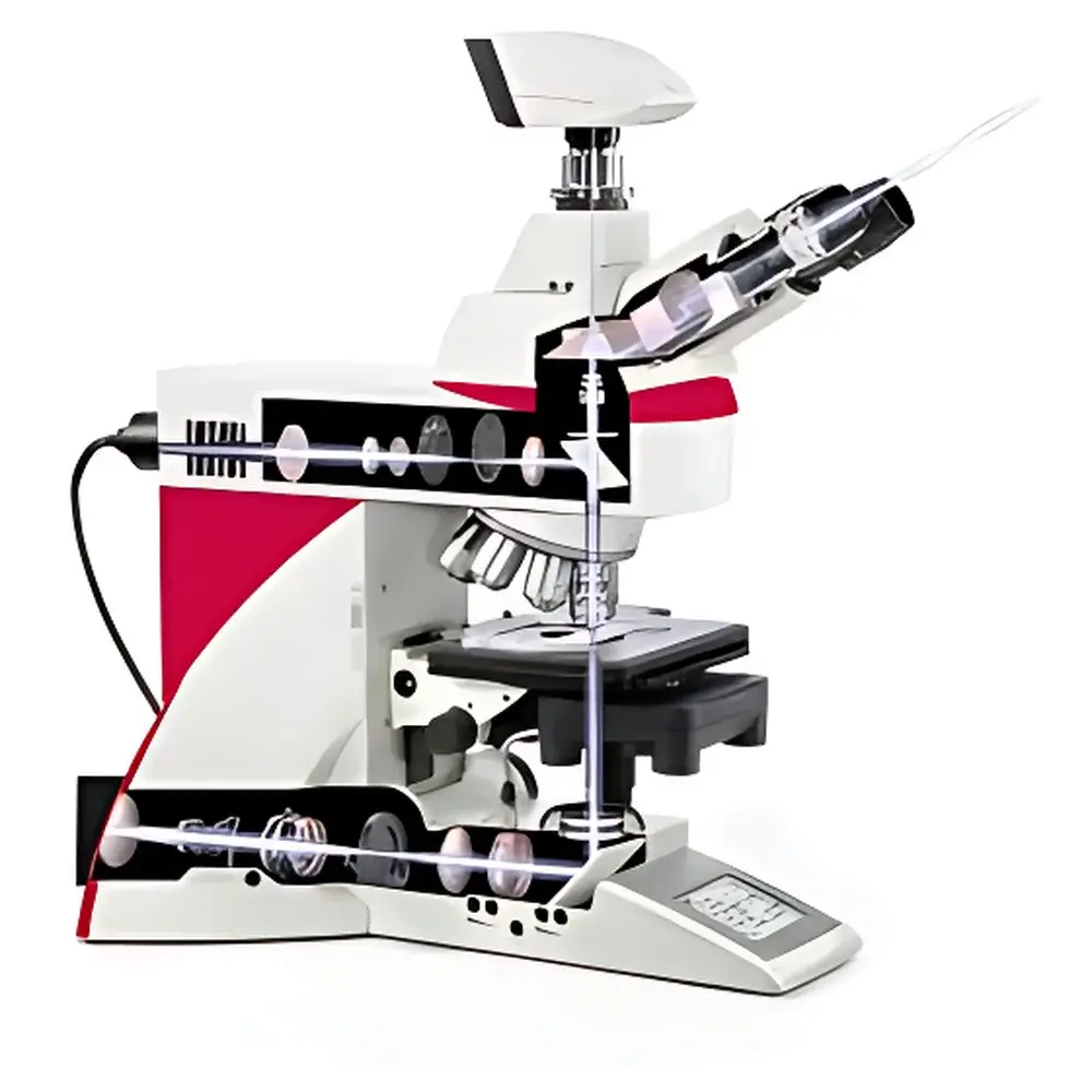
Leica DM6 B Upright Digital Research Microscope
| Brand | Leica |
|---|---|
| Origin | Germany |
| Model | Leica DM6 B |
| Illumination Options | Halogen or LED (with Constant Color Intensity Control, CCIC @ 3200 K) |
| Camera Port | 19-mm sCMOS-compatible |
| Objective Compatibility | Over 300 Leica objective lenses including 1.25× overview objective |
| Software Platform | Leica Application Suite X (LAS X) with Overview Navigation, Fluorescence Intensity Management (FIM), Excitation Manager (ExMan), and Köhler Illumination Management |
| Compliance | Designed for GLP/GMP environments |
| Automation Level | Fully motorized stage, focus, turret, condenser, and illumination |
Overview
The Leica DM6 B is a high-performance upright digital research microscope engineered for precision, reproducibility, and workflow efficiency in life science laboratories. Built on Leica Microsystems’ decades of optical expertise, the DM6 B employs advanced Köhler illumination architecture, motorized optical path management, and intelligent automation to deliver consistent, publication-grade imaging across brightfield, phase contrast, differential interference contrast (DIC), and fluorescence modalities. Its modular design integrates seamlessly with sCMOS cameras—specifically optimized via the 19-mm image port—to preserve full sensor resolution and quantum efficiency. The system operates on a rigid, thermally stable mechanical platform, minimizing drift during time-lapse or multi-position acquisitions. As a core instrument in academic, pharmaceutical, and clinical pathology settings, the DM6 B supports rigorous experimental protocols requiring traceable, repeatable imaging outcomes aligned with ISO/IEC 17025 and CLIA-compliant laboratory practices.
Key Features
- Fully motorized optical train: automated nosepiece, focus drive, XY stage, condenser height and centering, and illumination intensity—enabling one-button execution of complex acquisition routines.
- 19-mm sCMOS-optimized camera port: maintains full field-of-view coverage and pixel-level fidelity for high-resolution quantitative imaging without vignetting or magnification loss.
- Intelligent illumination management: halogen and LED options both supported; LED mode delivers stable 3200 K color temperature via Constant Color Intensity Control (CCIC), eliminating manual white balance recalibration between sessions.
- Fluorescence optimization suite: Fluorescence Intensity Management (FIM) dynamically adjusts exposure and lamp power per channel; Excitation Manager (ExMan) synchronizes filter cube selection with illumination source switching to minimize photobleaching.
- Objective ecosystem: compatible with >300 Leica HCX, PL FLUOTAR, and N PLAN objectives—including the 1.25× wide-field overview lens for rapid slide screening and region-of-interest (ROI) identification prior to high-magnification analysis.
- Software-driven navigation: LAS X software includes virtual slide overview mode, enabling mouse-controlled panning, zooming, and coordinate-based repositioning—critical for serial section alignment and multi-site time-lapse experiments.
Sample Compatibility & Compliance
The Leica DM6 B accommodates standard glass slides (1–3 mm thickness), petri dishes, multi-well plates, and custom specimen holders for live-cell imaging (when paired with environmental chambers). It supports all common histological preparations—including paraffin-embedded, frozen, resin-embedded, and immunolabeled sections—as well as unstained live specimens using phase contrast or DIC. For regulatory workflows, LAS X Enterprise Edition provides electronic signatures, role-based user permissions, and immutable audit trails compliant with FDA 21 CFR Part 11, EU Annex 11, and ISO 13485 quality management systems. Instrument configuration logs, acquisition metadata, and calibration records are embedded directly into exported image files (TIFF, ND2, or Leica’s native LIF format), ensuring full traceability from raw data to final publication figure.
Software & Data Management
Leica Application Suite X (LAS X) serves as the central interface for acquisition, analysis, and documentation. Its modular architecture allows labs to deploy only required modules—e.g., LAS X Core for basic imaging, LAS X Life Science for quantitative cell morphology, or LAS X Histology for tissue segmentation and annotation. All modules support batch processing, macro scripting (via Python API), and direct export to downstream platforms including ImageJ/Fiji, MATLAB, and commercial AI-based analysis tools. Metadata—including objective ID, exposure parameters, stage coordinates, and illumination settings—is automatically embedded in EXIF and TIFF tags. When connected to Leica’s LAS X Server, the system enables centralized storage, version-controlled project sharing, and remote monitoring—facilitating multi-user collaboration while maintaining data integrity under GLP/GMP audit conditions.
Applications
The DM6 B is routinely deployed in neurobiology for dendritic spine quantification, in cancer research for tumor margin assessment in frozen sections, in developmental biology for whole-embryo time-lapse imaging, and in clinical cytogenetics for karyotype analysis. Its phase contrast and DIC capabilities enable label-free visualization of organelle dynamics in primary hepatocytes or cardiomyocytes. In translational pathology, integration with Leica’s laser microdissection (LMD) systems permits precise isolation of morphologically defined cell populations—such as tumor-infiltrating lymphocytes—from H&E- or IHC-stained sections, followed by direct genomic or proteomic profiling. The microscope also supports correlative light-electron microscopy (CLEM) workflows when combined with Leica EM Cryo CLEM sample carriers.
FAQ
Is the Leica DM6 B compatible with third-party sCMOS cameras?
Yes—the 19-mm image port conforms to standard C-mount and F-mount mechanical interfaces; however, optimal performance (e.g., vignetting correction, pixel mapping, and hardware-triggered synchronization) is guaranteed only with Leica DFC7000 T, DFC9000 GT, or selected PCO and Hamamatsu models validated in LAS X.
Can the DM6 B be upgraded to support super-resolution techniques?
No—the DM6 B is not designed for STED, SIM, or PALM/STORM; those modalities require dedicated platforms such as the Leica STED CW or SR GSD 3D. However, it serves as an ideal pre-screening and validation tool for samples destined for super-resolution analysis.
Does the system support automated Z-stack acquisition with focus mapping?
Yes—LAS X includes autofocus algorithms (contrast-based and hardware-assisted) that generate focus maps across large fields, enabling reliable Z-series acquisition even on uneven or tilted specimens.
What maintenance is required for long-term calibration stability?
Annual verification of Köhler alignment, stage calibration, and objective encoding accuracy is recommended; Leica-certified service engineers perform traceable adjustments using NIST-traceable reference standards and issue ISO/IEC 17025-compliant calibration reports.
Is remote operation possible for core facility shared-resource environments?
Yes—via LAS X Remote Access (licensed option), users can control acquisition, adjust settings, and review images from any network-connected workstation without installing local software, provided authentication and encryption protocols meet institutional IT security policies.


