Examine R Confocal Raman Microscope
| Origin | Germany |
|---|---|
| Manufacturer Type | Authorized Distributor |
| Import Status | Imported |
| Model | Examine R |
| Pricing | Available Upon Request |
Overview
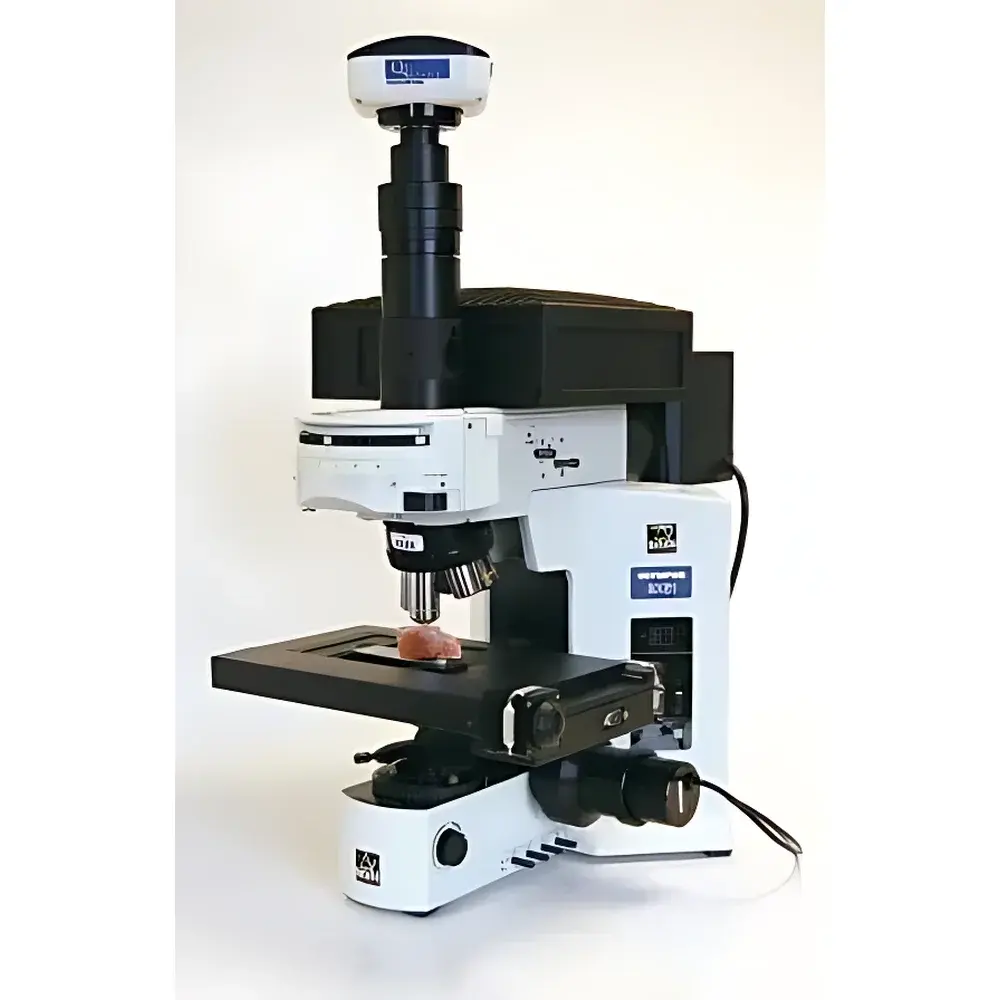
The Examine R Confocal Raman Microscope is a research-grade, modular confocal Raman imaging system engineered for high-spatial-resolution chemical characterization at the micro- and sub-micron scale. It integrates an Olympus upright or inverted optical microscope platform with a DeltaNu high-throughput, thermoelectrically cooled Raman spectrometer—enabling precise spectral acquisition across three excitation wavelengths (532 nm, 785 nm, and 1064 nm) without mechanical realignment of gratings, detectors, or laser paths. The system operates on confocal principle: spatial filtering via a pinhole aperture eliminates out-of-focus signal, delivering true depth-sectioning capability and diffraction-limited lateral resolution (~0.5 µm at 532 nm, ~0.65 µm at 785 nm, ~0.9 µm at 1064 nm under optimal conditions). This architecture supports non-destructive, label-free molecular fingerprinting in solid, liquid, and heterogeneous samples—making it indispensable for applications demanding both morphological context and vibrational spectroscopic specificity.
Key Features
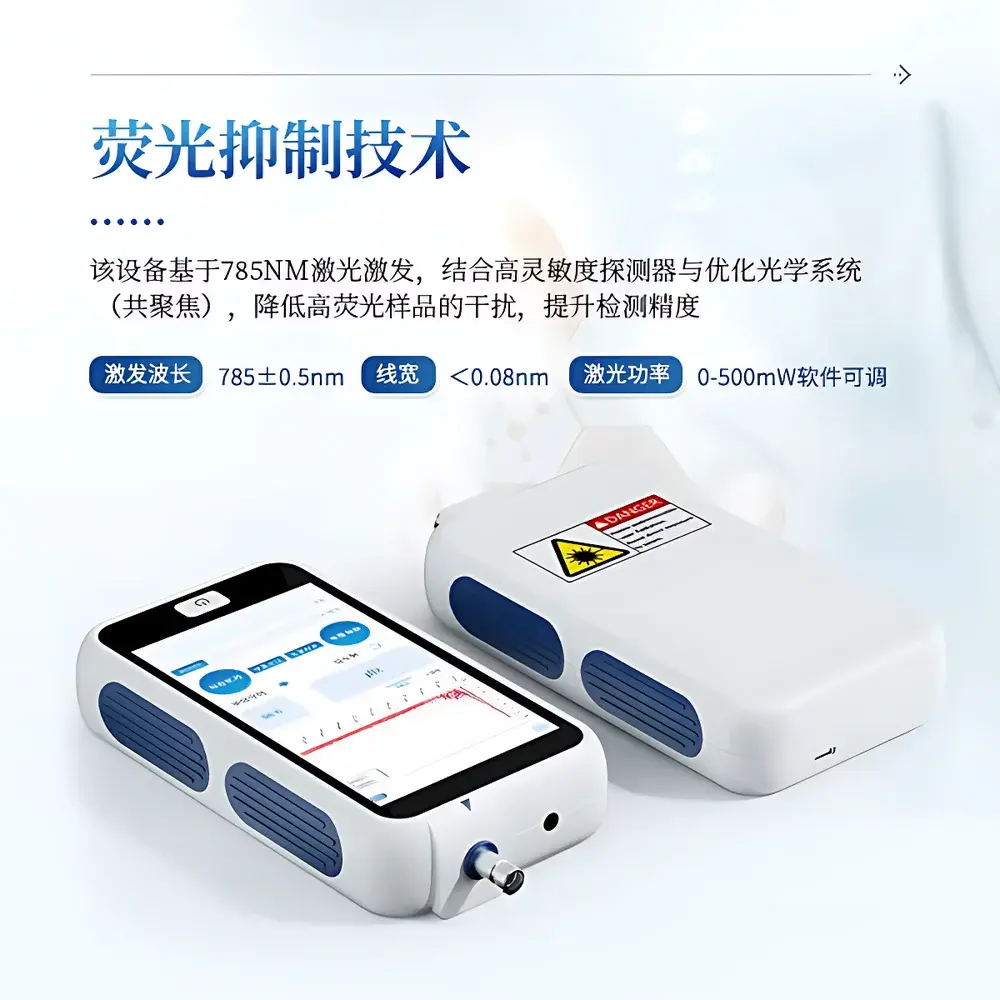
- Triple-wavelength laser compatibility: Interchangeable 532 nm, 785 nm, and 1064 nm diode laser modules—each with software-controlled power regulation (0–100% fine adjustment) to minimize photodamage in sensitive biological or polymeric specimens.
- High spectral fidelity: Fixed-grating, all-solid optical path ensures long-term alignment stability and reproducibility; no moving parts in the spectrometer core.
- Wide spectral coverage per acquisition: Up to 200–2000 cm⁻¹ (1064 nm, InGaAs detector), 100–2000 cm⁻¹ or 200–2000 cm⁻¹ (785 nm, NIR-optimized CCD), and 200–3400 cm⁻¹ (532 nm, deep-depletion CCD).
- Diffraction-limited microscopy: Olympus optical train delivers high NA objectives (up to 100×, 0.95 NA) for optimal light collection and spatial resolution.
- Motorized XYZ stage: High-precision, closed-loop piezo-driven stage enables automated Raman mapping with step sizes down to 100 nm; Z-stack acquisition supports 3D chemical reconstruction.
- Integrated visible-light imaging: High-resolution CMOS camera co-aligned with the Raman path provides real-time sample visualization, focus confirmation, and region-of-interest selection prior to spectral acquisition.
- Liquid-cell compatibility: Designed for in-situ Raman analysis in aqueous or organic solvent environments using standard capillary cells, flow cells, or custom sealed chambers.
Sample Compatibility & Compliance
The Examine R accommodates diverse sample types—including thin sections, polished wafers, geological thin sections, single cells, tissue slices, nanoparticles, polymer blends, and electrochemical interfaces—without requiring conductive coating or vacuum. Its open optical design permits integration with environmental stages (temperature-controlled, humidity-regulated, or gas-purged) and electrochemical cells. From a regulatory standpoint, the system supports GLP-compliant workflows: spectral metadata (laser wavelength, power, integration time, objective, grating, detector temperature) is embedded automatically in each spectrum file (e.g., .spa, .txt, or HDF5). Optional audit-trail logging and user-access controls align with FDA 21 CFR Part 11 readiness when paired with validated third-party LIMS or data management software.
Software & Data Management
Control and analysis are performed via DeltaNu’s SpectraWiz® software suite—a Windows-based application supporting instrument calibration, multi-point mapping, spectral library matching (with NIST, RRUFF, and custom databases), multivariate analysis (PCA, cluster analysis), and false-color Raman image generation. Raw data export is supported in ASCII, CSV, and industry-standard JCAMP-DX formats. For advanced quantification and batch processing, MATLAB and Python APIs (via DLL or REST interface) allow integration into automated analytical pipelines. All spectral acquisitions are timestamped and tagged with full hardware configuration logs, ensuring traceability required for ISO/IEC 17025-accredited laboratories.
Applications
- Life Sciences: Subcellular organelle identification, drug distribution in tissue, protein conformational changes, and label-free bacterial strain differentiation.
- Geosciences: Mineral phase mapping in thin sections, fluid inclusion analysis, carbon speciation in metamorphic rocks.
- Nanomaterials & Semiconductors: Strain mapping in SiGe heterostructures, defect identification in 2D materials (e.g., graphene layer count, MoS₂ crystallinity), and dopant distribution profiling.
- Forensics: Non-destructive identification of trace evidence—paint chips, fibers, explosives residues, and counterfeit pharmaceuticals—directly on substrates.
- Materials Chemistry: In-situ monitoring of polymer curing, battery electrode degradation, and catalytic surface reactions under controlled atmospheres.
FAQ
Is the Examine R compatible with third-party microscopes?
No—the system is factory-integrated with Olympus microscope optics and mechanically optimized for DeltaNu spectrometer coupling; retrofitting to non-Olympus platforms is not supported.
Can I perform time-resolved Raman measurements?
Yes—software-triggered acquisition sequences support kinetic studies with temporal resolution limited only by detector readout speed and laser pulse stability (down to ~10 ms per spectrum in rapid-scan mode).
What spectral calibration standards are included?
NIST-traceable silicon (520.7 cm⁻¹), cyclohexane (2847 cm⁻¹ C–H stretch), and neon emission lines are provided for daily wavelength and intensity calibration.
Does the system support polarization-resolved Raman?
Yes—optional motorized half-wave plate and polarizer modules enable polarization-dependent measurements for symmetry analysis and anisotropic material characterization.
Is remote operation possible?
Yes—network-enabled control allows secure remote access via VNC or RDP; full instrument functionality—including mapping and real-time imaging—is preserved over LAN/WAN connections.