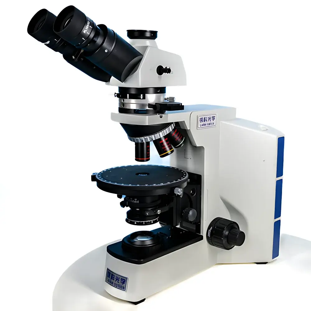
Lei-Tech LK-53 Research-Grade Upright Biological Fluorescence Microscope
| Brand | Lei-Tech |
|---|---|
| Origin | Tianjin, China |
| Manufacturer Type | Direct Manufacturer |
| Product Category | Domestic |
| Model | LK-53 |
| Instrument Type | Upright Microscope |
| Eyepiece Configuration | Trinocular |
| Optical System | Infinity-Corrected Chromatic Aberration-Corrected (UISC) |
| Total Magnification Range | 40×–1000× (with 4×, 10×, 20×, 40×, 100× objectives) |
| Illumination | LED Transmitted Light + Four-Channel LED Epifluorescence (B: 470 nm, G: 560 nm, UV2: 385 nm) |
| Objective NA | Up to 1.4 (Plan Apochromat Fluorescence Objectives) |
| Eyepieces | Widefield PLN10×/22 mm, High-Eyepoint, Optional Reticle |
| Nosepiece | Internal-Positioning Five-Objectives Turret |
| Focus Mechanism | Coaxial Coarse/Fine Adjustment, 30 mm Coarse Travel, 2 µm Fine Step Resolution |
| Stage | Dual-Layer Mechanical X-Y Translation Platform |
| Condenser | Swing-Out Achromatic Condenser, NA 1.2 / 0.22 |
| Camera Interface | C-mount, 20 MP Sony Exmor CMOS Sensor (5440 × 3648), Active Thermoelectric Cooling (ΔT = −40°C vs. ambient), Anti-Fog Optics |
| Image Processing | Real-Time HDR (DHR), Z-Stack Depth Fusion, Multi-Field Mosaic Stitching |
| Filter Wheel | Precision Disk-Based Six-Position Fluorescence Filter Holder |
| Power Supply | Universal Input 100–240 V AC, Digital Control with Usage Timer & Current Monitoring |
Overview
The Lei-Tech LK-53 is a research-grade upright biological fluorescence microscope engineered for high-fidelity multimodal imaging in academic laboratories, clinical pathology units, and industrial R&D environments. Built upon an infinity-corrected optical architecture (UISC — Universal Infinity Space Correction), the LK-53 eliminates field curvature and chromatic aberration across the full magnification range (40× to 1000×), enabling quantitative consistency between brightfield, phase contrast, polarized light, darkfield, and epifluorescence observation modes. Its dual-path illumination system integrates a stabilized high-CRI white LED for transmitted-light applications and a four-channel LED epifluorescence excitation module—featuring precisely centered 470 nm (blue), 560 nm (green), and 385 nm (UV2 long-pass) bands—designed to minimize thermal drift and spectral crosstalk. The system’s optical train incorporates Plan Apochromat fluorescence objectives with numerical apertures up to 1.4, multi-layer broadband anti-reflective coatings optimized for UV-to-visible transmission (>92% at 385 nm), and fluorophore-specific dichroic beam splitters with <1% autofluorescence background. This architecture ensures high signal-to-noise ratio, submicron lateral resolution, and reproducible photometric linearity—critical for longitudinal cell tracking, colocalization analysis, and semi-quantitative fluorescence intensity profiling.
Key Features
- Infinity-corrected UISC optical pathway with parfocal, parcentric alignment across all objectives and modules
- Trinocular observation head with 30° inclination, 360° rotatable eyepiece tube, and adjustable interpupillary distance (54–75 mm)
- Five-position internal-locking objective turret supporting dry and oil-immersion objectives (4×, 10×, 20×, 40×, 100×)
- Coaxial coarse/fine focusing mechanism with 30 mm vertical travel and 2 µm fine-step resolution; includes upper-limit stop and tension adjustment
- Swing-out achromatic condenser (NA 1.2 / 0.22) with centerable aperture and field diaphragms for Köhler illumination optimization
- Six-position precision disk-based fluorescence filter changer—mechanically indexed for repeatable alignment and minimal vibration-induced misregistration
- Dedicated thermoelectrically cooled 20 MP Sony Exmor CMOS camera (5440 × 3648 pixels), operating at −40°C below ambient with anti-fog optical housing
- Real-time digital image processing suite: Dynamic High Dynamic Range (DHR) tone mapping, Z-stack depth fusion, and multi-field mosaic stitching
- Digital power management system with runtime counter, current monitoring, and LED usage diagnostics—compatible with GLP-compliant instrument logbook protocols
Sample Compatibility & Compliance
The LK-53 accommodates standard glass microscopy slides (26 × 76 mm), coverslips (No. 1.5, 0.17 mm thickness), and live-cell chambers (e.g., 35 mm Petri dishes, Ibidi µ-Slides). Its modular design supports routine histopathology sections, frozen tissue slices, cytospin preparations, and adherent or suspension cultures under controlled environmental enclosures (optional). The system conforms to ISO 10993-5 (biocompatibility of optical components in contact with specimens), IEC 61000-4-2/3/4 (EMC immunity), and CE marking requirements for laboratory equipment. All fluorescence modules comply with EN 62471 (photobiological safety), and the LED excitation sources are certified Class 1M under IEC 60825-1. While not FDA 510(k)-cleared as a diagnostic device, the LK-53 meets ASTM E2877-22 criteria for fluorescence microscope performance verification and supports audit-ready documentation for ISO/IEC 17025-accredited testing labs.
Software & Data Management
The LK-53 operates with Lei-Tech’s proprietary Imaging Suite v4.2—a Windows-based application supporting DICOM, TIFF, and OME-TIFF export formats. It implements role-based user authentication, electronic signature workflows, and time-stamped audit trails compliant with FDA 21 CFR Part 11 Annex 11 requirements. Image metadata (objective ID, exposure time, gain, LED intensity, filter position, stage coordinates) is embedded automatically. Batch acquisition scripts enable automated multi-channel, multi-z, multi-position experiments with configurable dwell times and focus correction loops. Raw sensor data is preserved without lossy compression; processed images retain non-destructive layer history. Exported datasets are compatible with open-source platforms including Fiji/ImageJ, QuPath, and CellProfiler via standardized metadata headers.
Applications
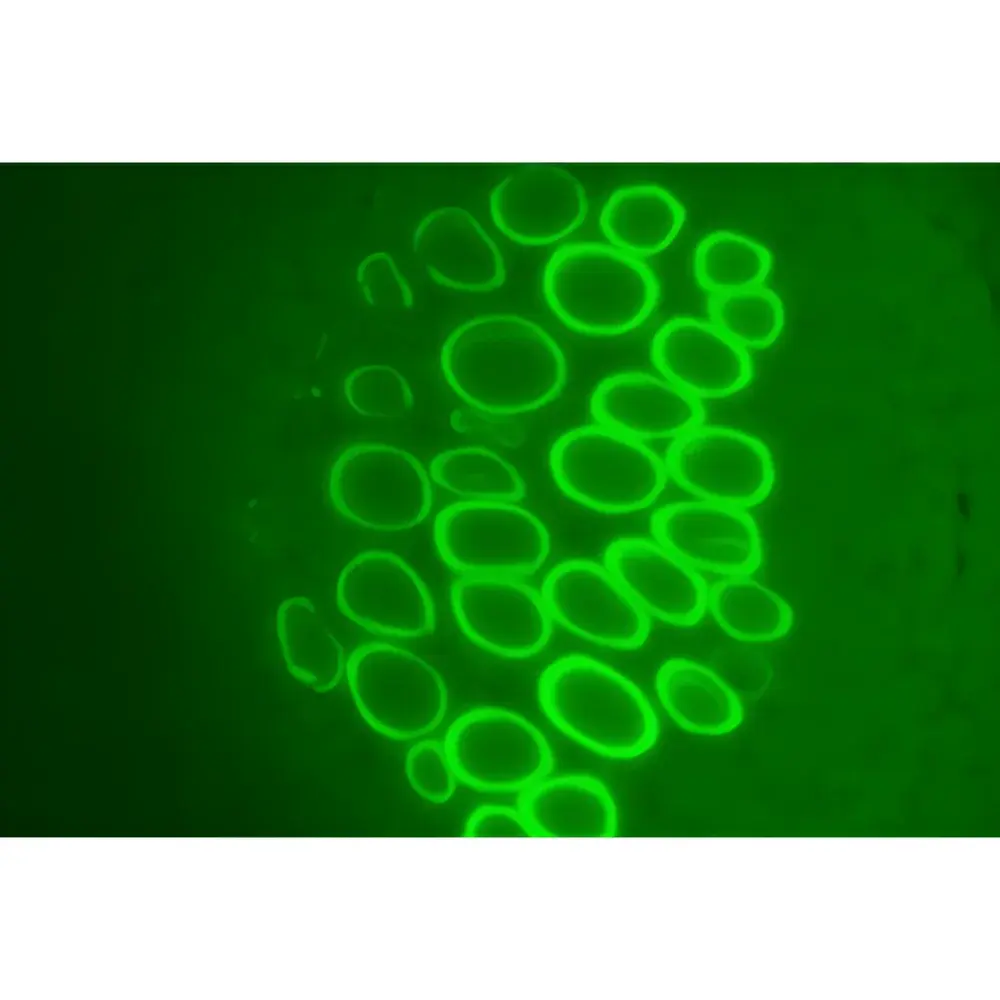
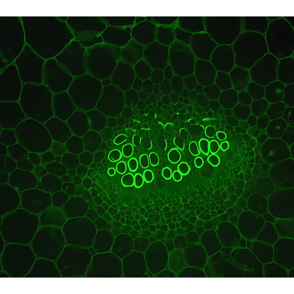
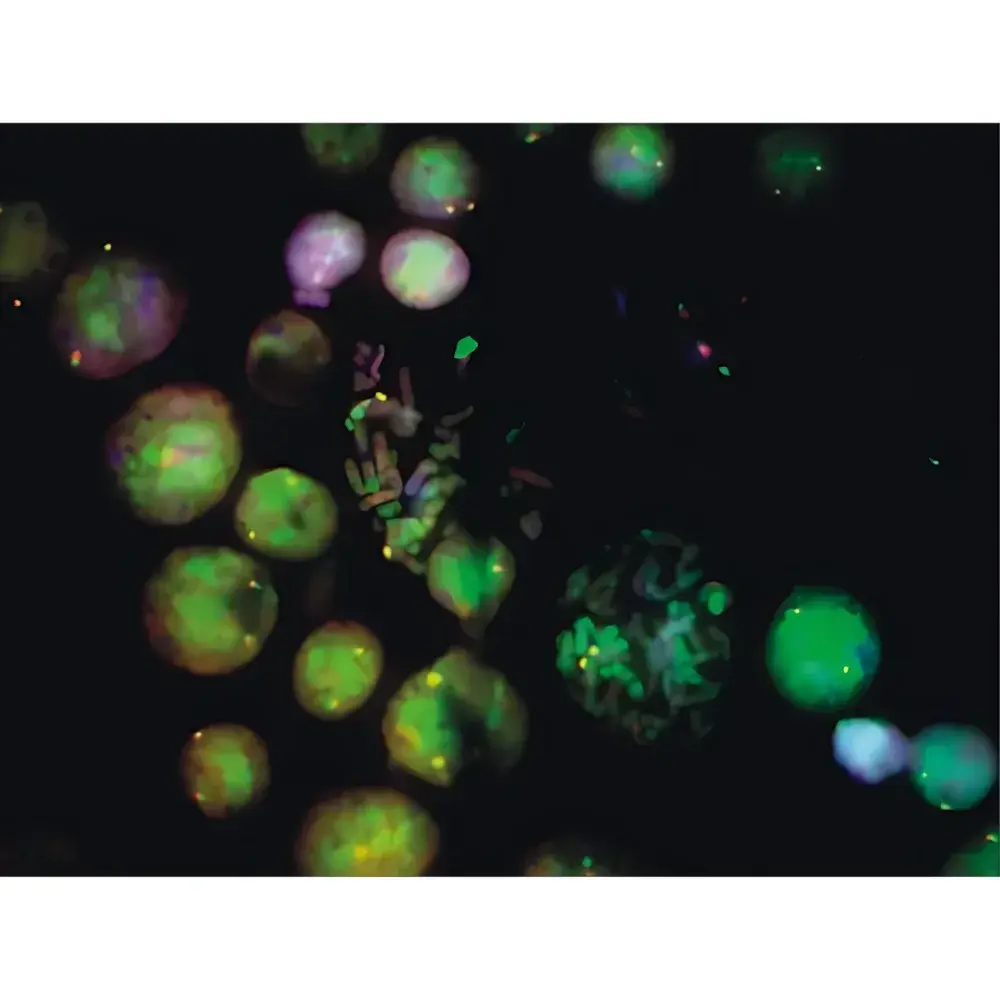
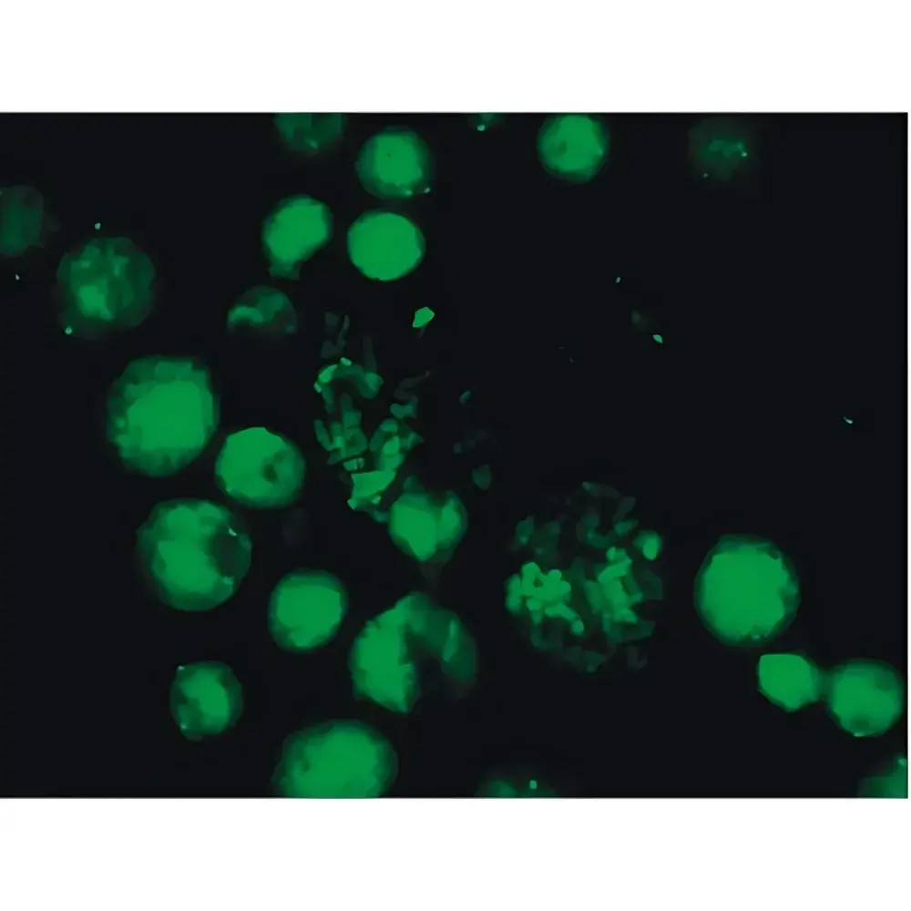
The LK-53 serves as a primary imaging platform for immunofluorescence localization (e.g., nuclear vs. cytoplasmic marker colocalization), mitotic staging in live or fixed cells, fluorescent protein expression quantification (GFP, mCherry, tdTomato), calcium flux imaging using Fluo-4 or Fura-2 dyes, and routine H&E-adjacent fluorescence validation in diagnostic pathology. Its phase contrast and brightfield capabilities support unstained live-cell morphology assessment, while polarized light mode enables birefringence analysis of collagen, starch granules, or liquid crystals. In teaching laboratories, its intuitive interface and modular configuration facilitate hands-on instruction in optical principles, specimen preparation, and digital image analysis fundamentals. Industrial users deploy it for QC of antibody-conjugated nanoparticles, microcarrier-based bioreactor monitoring, and semiconductor wafer defect inspection requiring high-contrast visible-light resolution.
FAQ
Does the LK-53 support oil-immersion objectives?
Yes—it is fully compatible with standard 100× NA 1.4 oil-immersion Plan Apochromat objectives, including third-party lenses meeting RMS thread specifications.
Is the camera sensor cooled during extended acquisitions?
Yes—the integrated Peltier cooling system maintains the Sony Exmor CMOS sensor at a stable −40°C delta relative to ambient temperature, suppressing dark current noise even during 30-minute time-lapse sequences.
Can the system be validated for ISO/IEC 17025 compliance?
Yes—Lei-Tech provides a traceable calibration certificate (NIST-traceable stage micrometer and neutral density filters), IQ/OQ documentation templates, and on-site performance qualification support.
What fluorescence filter sets are included by default?
The base configuration includes DAPI/FITC/TRITC/Cy5-compatible sets with hard-coated, edge-steepness-optimized dichroics and bandpass filters; custom OEM sets (e.g., GFP/mCherry, Hoechst/CFP/YFP) are available upon request.
Is remote operation supported?
Yes—the Imaging Suite supports secure LAN/WAN access via TLS-encrypted API endpoints, enabling centralized control from core facility workstations or cloud-managed lab networks.


