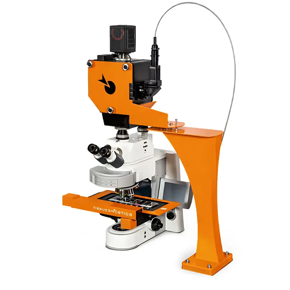
TissueGnostics TISSUEFAXS Q+ High-Resolution Multidimensional Whole-Slide Spatial Phenotyping and Single-Cell Quantification System
| Brand | TissueGnostics |
|---|---|
| Country of Origin | Austria |
| Manufacturer Type | Original Equipment Manufacturer (OEM) |
| Import Status | Imported |
| Model | TISSUEFAXS Q+ |
| Price Range | USD 420,000 – 700,000 |
Overview
The TissueGnostics TISSUEFAXS Q+ is an integrated high-resolution whole-slide imaging and spatial phenotyping platform engineered for quantitative, in situ single-cell analysis across intact tissue sections. It operates on the principle of automated, seamless panoramic scanning combined with multispectral fluorescence and brightfield acquisition—enabling pixel-accurate registration of morphological, molecular, and spatial context at cellular resolution across entire glass slides (up to 150 × 150 mm). Unlike conventional microscopy systems limited to field-of-view–constrained analysis, the TISSUEFAXS Q+ captures comprehensive spatial architecture by stitching high-magnification image tiles with submicron positional fidelity. Its optical architecture incorporates a motorized precision stage, high-stability objective turret (supporting 2x–63x objectives), and a scientific CMOS sensor optimized for low-noise, high-dynamic-range acquisition. The system is fundamentally designed for spatial biology workflows requiring rigorous quantification of cell phenotype, density, proximity, and neighborhood topology within native tissue microenvironments.
Key Features
- Automated whole-slide scanning with real-time autofocus and drift compensation for consistent Z-stack acquisition across heterogeneous tissue thicknesses
- Modular illumination: Independently controllable multi-channel LED light sources (365–740 nm) with programmable intensity and exposure timing to minimize photobleaching during sequential channel acquisition
- Spinning disk confocal module (optional) enabling optical sectioning at 20× magnification—validated for mouse whole-brain coronal sections with axial resolution <1.2 µm
- Integrated high-throughput slide loader supporting up to 200 standard glass slides per unattended run, compliant with ANSI/SBS microplate footprint standards
- Dual-mode imaging engine: Simultaneous brightfield (H&E, DAB, Masson’s trichrome) and widefield fluorescence (DAPI, FITC, Cy3, Cy5, AF647) acquisition with spectral unmixing capability
- Hardware-synchronized stage motion and camera triggering ensuring sub-pixel image alignment and zero stitching artifacts
Sample Compatibility & Compliance
The TISSUEFAXS Q+ accommodates standard FFPE and frozen tissue sections (4–50 µm thickness), cytospin preparations, and tissue microarrays (TMAs) mounted on standard microscope slides. It supports common histological staining protocols including IHC, IF, multiplexed immunofluorescence (up to 7-plex with spectral separation), and chromogenic in situ hybridization (CISH). All hardware and software components comply with ISO 13485:2016 (medical device quality management) and CE IVD marking requirements. Data handling adheres to ALCOA+ principles (Attributable, Legible, Contemporaneous, Original, Accurate, Complete, Consistent, Enduring, Available) and supports audit trails required under FDA 21 CFR Part 11 for regulated preclinical research environments.
Software & Data Management
Acquisition and analysis are unified under TissueStudio 5.x—a validated, modular software suite featuring GPU-accelerated image processing pipelines. Core modules include: (1) TissueAlign for nonlinear distortion correction and multi-layer registration; (2) TissueQuest for AI-assisted cell segmentation (U-Net backbone trained on >10⁶ annotated nuclei); (3) StrataQuest for spatial statistics including nearest-neighbor distance mapping, Ripley’s K-function analysis, and graph-based neighborhood clustering. All processed datasets export FAIR-compliant metadata (MIAME/MINSEQE extensions), support OMERO interoperability, and integrate natively with downstream platforms such as QuPath, HALO, and R/Bioconductor via standardized OME-TIFF and GeoMx-compatible JSON formats. Audit logs record user actions, parameter changes, and processing history for GLP/GMP traceability.
Applications
- Translational biomarker discovery: Quantifying PD-L1⁺/CD8⁺ T-cell spatial co-localization in tumor immune microenvironments
- Neuroanatomical mapping: Whole-brain cell-type distribution profiling in transgenic mouse models using multiplexed IF
- TMA validation studies: High-throughput phenotypic scoring across hundreds of clinical cohorts with batch-corrected normalization
- Drug mechanism-of-action analysis: Measuring treatment-induced shifts in stromal–epithelial interaction metrics (e.g., fibroblast–tumor cell proximity entropy)
- Spatial transcriptomics correlation: Anchoring Visium or Xenium-derived gene expression hotspots to protein-level phenotypes at single-cell resolution
FAQ
What tissue section thicknesses are supported without compromising focus stability?
Standard protocols validate robust performance from 4 µm (FFPE) to 50 µm (vibratome-cut frozen sections), with adaptive Z-stack depth optimization enabled per region-of-interest.
Does the system support time-lapse imaging of live tissue cultures?
No—the TISSUEFAXS Q+ is optimized for fixed-tissue analysis; live-cell dynamics require dedicated incubator-integrated systems not part of this platform’s design scope.
Can third-party antibodies or custom dyes be used without protocol recalibration?
Yes—spectral library customization is supported via user-defined reference spectra; no hardware reconfiguration is required for new fluorophore integration.
Is cloud-based data storage and remote collaboration supported out-of-the-box?
Local deployment is standard; optional TissueCloud Connect module provides encrypted HTTPS API access, role-based permissions, and DICOM-SR export for PACS integration.
How is calibration traceability maintained across instrument lifetime?
NIST-traceable stage calibration slides and LED intensity reference standards are included; annual verification reports align with ISO/IEC 17025 laboratory accreditation requirements.


