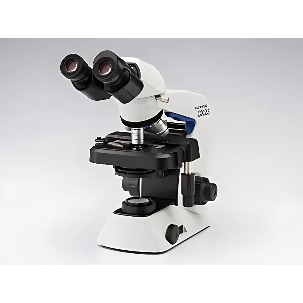


Olympus CX23 Biological Microscope
| Brand | Olympus |
|---|---|
| Origin | Japan |
| Model | CX23 |
| Type | Upright Biological Microscope |
| Optical System | Infinity-Corrected |
| Illumination | Integrated 0.5W LED Transmitted Light Source |
| Focus Mechanism | Coaxial coarse/fine focus with 15 mm coarse travel per revolution, 2.5 µm fine focus increment |
| Stage | Steel-wire driven mechanical stage (174 × 89 mm), 76 × 30 mm travel range |
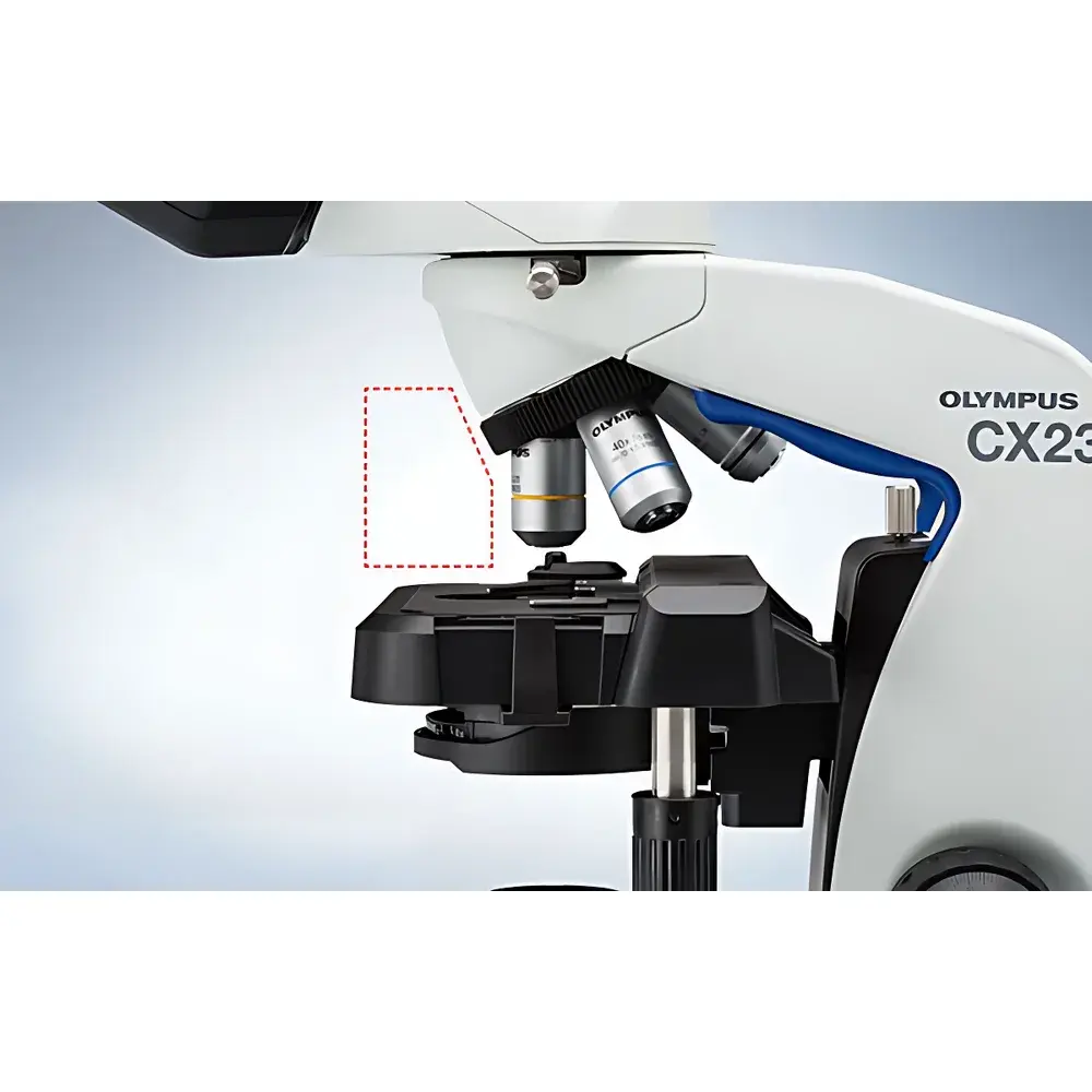
| Objective Turret | Fixed inward-swinging 4-position nosepiece |
| Objectives | Plan Achromat 4× (NA 0.10, WD 27.8 mm), 10× (NA 0.25, WD 8.0 mm), 40× (NA 0.65, WD 0.6 mm), 100× Oil (NA 1.25, WD 0.13 mm) |
| Eyepieces | Wide-field 10× (FN 20), optional 15× (FN 12), all anti-fungal coated |
| Interpupillary Distance | 48–75 mm |
| Eye-point Adjustment | 370.0–432.9 mm |
| Condenser | Abbe condenser (NA 1.25 with oil immersion), built-in iris diaphragm |
| Power Supply | AC 100–240 V, 50/60 Hz, <2 W consumption |
| Dimensions (W×D×H) | 198 × 398 × 386 mm (binocular), 198 × 398 × 430 mm (trinocular) |
| Weight | ~5.9 kg (binocular), ~6.5 kg (trinocular) |
| Compliance | CE, RoHS, ISO 9001-manufactured design |
Overview
The Olympus CX23 is a robust, entry-level upright biological microscope engineered for routine laboratory use in academic teaching, clinical diagnostics, and quality control environments. Built upon Olympus’ decades of optical engineering heritage, the CX23 employs an infinity-corrected optical system to deliver consistent image fidelity across the full field of view. Its core imaging principle relies on transmitted brightfield illumination—optimized via a thermally stable, low-power 0.5W LED source—to support standard histological preparations including H&E-, Giemsa-, and Gram-stained specimens. The system’s mechanical stability, ergonomic accessibility, and anti-fungal optical component treatment make it particularly suited for high-humidity laboratories and shared-use facilities where long-term reliability and minimal maintenance are critical operational requirements.
Key Features
- Infinity-corrected optical architecture: Ensures chromatic and spherical aberration correction across magnifications, enabling sharp, flat-field imaging with plan achromat objectives (4× to 100× oil).
- Dual-purpose mechanical stage: Steel-wire driven, fixed-stage design (174 × 89 mm) with 76 mm × 30 mm travel range and integrated specimen clips; includes engraved positioning scale for repeatable sample navigation.
- Ergonomic focusing system: Coaxial coarse/fine focus knobs with torque-limiting mechanism to prevent gear damage during over-rotation; fine focus resolution of 2.5 µm per division supports precise Z-axis localization.
- Inward-swinging 4-position objective turret: Provides unobstructed access above the stage—facilitating rapid oil immersion application and safe handling of thick or multi-layered specimens.
- Anti-fungal optical train: All oculars, objectives, and observation tubes undergo proprietary anti-microbial coating treatment—validated under ISO 846 (plastics — evaluation of resistance to microbial growth)—to inhibit fungal colonization in tropical or humid operational climates.
- LED illumination system: 20,000-hour rated, color-temperature-stabilized blue-enhanced LED (peak ~455 nm) optimized for contrast retention in hematoxylin-eosin stained tissues; power consumption remains below 2 W, supporting energy-efficient lab operations.
Sample Compatibility & Compliance
The CX23 accommodates standard glass microscope slides (76 × 26 mm) and cover slips (0.13–0.17 mm thickness) across all magnification ranges. Its Abbe condenser (NA 1.25) supports both dry and oil-immersion protocols, with calibrated iris diaphragm control for Köhler illumination alignment. The system complies with IEC 61000-6-3 (EMC emission standards) and IEC 61000-6-1 (immunity), carries CE marking per Directive 2014/30/EU (EMC) and 2014/35/EU (LVD), and adheres to RoHS 2011/65/EU material restrictions. While not FDA 510(k)-cleared as a medical device, its optical performance meets ASTM E2868–22 (Standard Guide for Microscopical Examination of Biological Specimens) criteria for educational and non-diagnostic investigative use.
Software & Data Management
The CX23 is a hardware-only platform with no embedded digital imaging module. However, its trinocular configuration (100/0 or 0/100 beam splitter) enables seamless integration with third-party C-mount cameras (e.g., USB3.0 CMOS sensors) and widely adopted microscopy software suites—including Olympus cellSens Entry (via optional adapter), ImageJ/Fiji (open-source), and HALCON-based custom acquisition pipelines. All digital outputs retain native 1:1 pixel mapping without interpolation artifacts when paired with ≤5 MP sensors. Audit trail functionality (e.g., timestamped image metadata, user ID tagging) is fully supported through external software compliant with 21 CFR Part 11 when deployed in GLP/GMP-aligned workflows.
Applications
- Undergraduate and graduate life science instruction: Cell morphology, mitotic staging, protozoan motility, and basic histology.
- Clinical screening labs: Peripheral blood smear review, urinalysis sediment examination, and fungal element identification (e.g., Candida, Aspergillus hyphae).
- Pharmaceutical QC: Visual inspection of sterile filtration validation samples, excipient particle distribution, and tablet coating uniformity.
- Agricultural pathology: Plant tissue section analysis, nematode identification, and seed viability assessment.
- Environmental microbiology: Activated sludge floc structure evaluation and biofilm thickness estimation in wastewater monitoring.
FAQ
Is the CX23 compatible with digital camera systems?
Yes—the trinocular version features a C-mount port with 100/0 or 0/100 light path selection, supporting standard 1/2″ or 1/3″ sensor cameras.
Does the CX23 meet ISO 13485 or FDA 21 CFR Part 11 requirements?
The microscope itself is not certified to these standards; however, when integrated with validated third-party imaging software and documented SOPs, it may be included in Part 11–compliant workflows.
Can oil immersion objectives be used without risk of stage collision?
Yes—the inward-swinging nosepiece design increases vertical clearance above the stage, minimizing contact risk during 100× oil objective engagement.
What is the expected lifetime of the LED illumination source?
Rated for 20,000 hours at nominal output—equivalent to >10 years of typical academic lab usage (4 hrs/day, 250 days/year).
Is service calibration available outside Japan?
Olympus-certified service partners in over 40 countries provide optical alignment verification, focus mechanism recalibration, and LED intensity profiling per Olympus Technical Bulletin TB-CX23-Rev.D.


