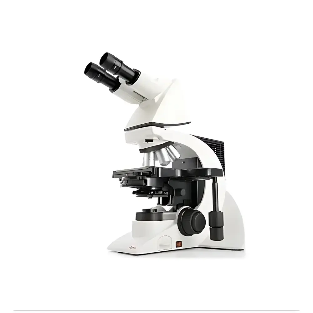
Leica DM2000 Biological Microscope
| Origin | Germany |
|---|---|
| Manufacturer Type | Authorized Distributor |
| Origin Category | Imported |
| Model | DM2000 |
| Pricing | Upon Request |
Overview
The Leica DM2000 is a modular, research-grade upright biological microscope engineered for high-precision transmitted-light imaging in demanding life science applications. Built on Leica Microsystems’ proven optical architecture, it employs Köhler illumination with a 12 V / 30 W halogen lamp to deliver uniform, stable, and glare-free illumination—essential for quantitative brightfield, darkfield, phase contrast, polarized light, and fluorescence microscopy. Its rigid mechanical design minimizes vibration-induced drift, while the coaxial focus mechanism ensures precise Z-axis control with fine and coarse focusing integrated into a single ergonomic axis. Designed for long-term reliability and user-specific adaptability, the DM2000 serves as a foundational platform in academic laboratories, clinical pathology departments, and quality-controlled diagnostic environments where reproducibility, regulatory traceability, and multi-technique flexibility are critical.
Key Features
- Modular optical platform supporting simultaneous integration of up to five contrast methods: brightfield, darkfield, phase contrast, polarization, and fluorescence
- Ergonomic, height-adjustable focus knobs with patented fingertip positioning—designed for operator-specific hand size and posture to reduce repetitive strain during extended use
- 7-position objective turret enabling seamless switching between Leica’s full range of plan achromat, plan semi-apochromat, and plan apochromat objectives (e.g., 4×, 10×, 20×, 40×, 63×, 100× oil)
- 5-position fluorescence filter cube slider with Leica’s Zero-Pixel-Shift technology—ensuring pixel-perfect registration across excitation/emission channels and eliminating image misalignment during multi-channel acquisition
- Binocular or trinocular observation tube options; compatible with 10× wide-field eyepieces (20–25 mm field number) and optional 12.5×, 15×, or 20× magnification eyepieces
- Integrated mechanical stage with low-profile X-Y controls aligned coaxially with focus knobs—optimized for coordinated manipulation and fatigue reduction during serial section analysis or cell tracking
- Quick-access drawer-style lamp housing for tool-free 30 W halogen bulb replacement—minimizing instrument downtime in high-throughput lab environments
Sample Compatibility & Compliance
The DM2000 accommodates standard 1 mm thick glass slides and coverslips (0.13–0.17 mm), supporting routine histological sections, cytology smears, live-cell monolayers (with appropriate environmental chambers), and fixed immunofluorescent preparations. Its optical path is calibrated to ISO 8578 (microscope mechanical tube length) and conforms to DIN/ISO 10934-1 for objective labeling and parfocality. The system supports GLP/GMP-aligned workflows when paired with validated digital imaging software: audit trails, user access control, and electronic signatures can be implemented per FDA 21 CFR Part 11 requirements. All fluorescence components comply with IEC 61000-4-3 (EMC immunity) and IEC 62471 (photobiological safety classification for UV/blue excitation sources).
Software & Data Management
The DM2000 interfaces seamlessly with Leica Application Suite (LAS X) and third-party imaging platforms including NIS-Elements, MetaMorph, and ImageJ via standard USB 3.0 or GigE Vision protocols. LAS X provides full hardware control—including motorized filter selection, exposure timing, Z-stack acquisition, and multi-channel overlay—with built-in calibration management for pixel size, intensity linearity, and color balance. Raw image metadata (objective ID, magnification, illumination mode, exposure time, camera gain) is embedded in TIFF/OME-TIFF headers, ensuring FAIR (Findable, Accessible, Interoperable, Reusable) data principles. Optional modules enable DICOM export for PACS integration in clinical pathology settings and CSV-based morphometric reporting compliant with CAP and CLIA documentation standards.
Applications
- Diagnostic histopathology and cytopathology in accredited medical laboratories
- Cell morphology assessment in stem cell differentiation and toxicology screening
- Immunofluorescence co-localization studies requiring sub-pixel registration accuracy
- Teaching laboratories requiring robust, serviceable instrumentation with multi-user configurability
- Quality assurance of tissue culture substrates and biomaterial coatings
- Forensic fiber and pollen analysis using polarized light and differential interference contrast
FAQ
Is the DM2000 compatible with LED illumination upgrades?
Yes—Leica offers the DM2000 LED retrofit kit (part no. 11506018), which replaces the halogen module with a thermally stabilized 3W white LED source delivering >12,000 hours lifetime and CIE 1931 chromaticity stability within ±0.002 Δu’v’.
Can the DM2000 support motorized components?
While the base DM2000 is manually operated, its optical train and mounting interface are fully compatible with Leica’s Motorized Focus Module (MFM) and Motorized Filter Wheel (MFW)—enabling automation-ready expansion without structural modification.
What objective correction levels are supported?
The system accepts all Leica plan-corrected objectives: Plan Achromat (PA), Plan Semi-Apochromat (PL FLUOTAR), and Plan Apochromat (HCX PL APO), with cover-slip correction collars adjustable from 0.13 to 0.23 mm.
Does the DM2000 meet ISO 9001-certified manufacturing standards?
All Leica DM-series microscopes are assembled and tested at the Wetzlar, Germany facility under ISO 9001:2015 and ISO 13485:2016 certified quality management systems, with full traceability of optical components and mechanical assemblies.
Related Products


