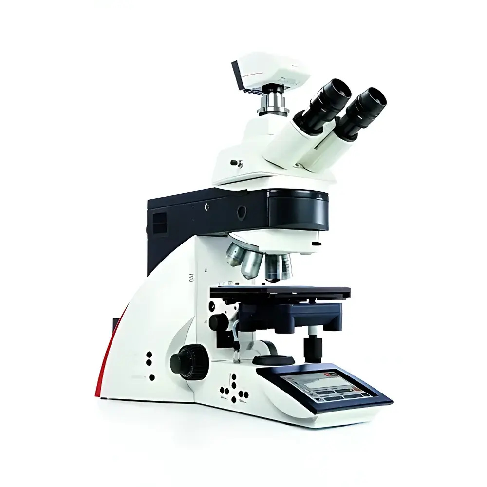
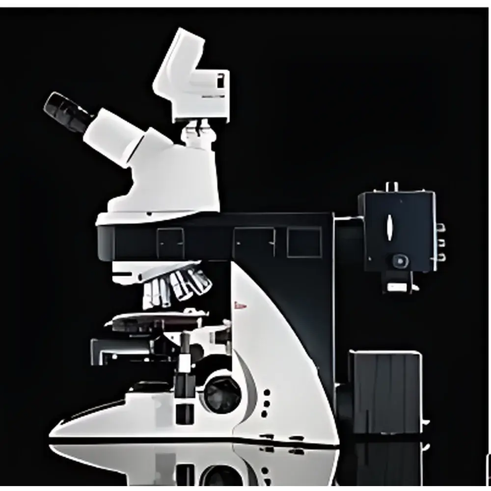
Leica DM5000B Biological Microscope
| Brand | Leica |
|---|---|
| Origin | Germany |
| Model | DM5000B |
| Optical Path | Infinity-Corrected |
| Illumination | 12 V / 100 W Halogen Transmitted-Light Source |
| Objective Turret | Motorized 7-Position Encoded Nosepiece |
| Focus Drive | Manual Z-Axis with Ergonomic Coaxial Knobs |
| Contrast Methods | Brightfield, Darkfield, Phase Contrast, Polarization, Differential Interference Contrast (DIC), Fluorescence |
| Fluorescence Module | Motorized 5-Position or 8-Position Filter Cube Turret with Fluorescence Intensity Manager (FIM) |
| Eyepiece | Widefield 10× (25 mm Field Number), Compatible with Optional Multi-Magnification Eyepieces |
| Observation Tube | Binocular or Trinocular |
| Software Integration | Leica Application Suite (LAS) X, Fully Compatible with Third-Party Imaging & Analysis Platforms |
| Compliance | Designed and Manufactured in Accordance with ISO 9001 and IEC 61000-6-3 Electromagnetic Compatibility Standards |
Overview
The Leica DM5000B is a high-performance, semi-automated upright biological microscope engineered for rigorous life science research environments—particularly live-cell imaging, tissue culture analysis, and morphological assessment of fixed or living specimens. Built upon Leica Microsystems’ proven infinity-corrected optical architecture, the DM5000B delivers diffraction-limited resolution, chromatic fidelity, and long-term mechanical stability across all contrast modalities. Its core design integrates motorized encoding and precision mechanics to ensure repeatable, operator-independent acquisition conditions—a critical requirement for longitudinal studies, regulatory-compliant documentation, and multi-user laboratory workflows. The system supports full transmittance-based imaging—including Leica’s proprietary automated Differential Interference Contrast (DIC)—and is optimized for integration with high-sensitivity sCMOS or EMCCD cameras, enabling quantitative fluorescence intensity profiling and time-lapse volumetric reconstruction.
Key Features
- Motorized 7-position encoded objective nosepiece with position memory and automatic magnification recognition—ensures accurate calibration traceability for image scaling and metadata embedding.
- Manual coaxial focus drive with dual-speed fine/coarse adjustment and ergonomic torque tuning—optimized for extended manual operation while maintaining sub-micron repeatability.
- Integrated Leica Fluorescence Intensity Manager (FIM)—dynamically regulates lamp output and filter positioning to maintain consistent photon flux across sequential acquisitions, minimizing phototoxicity and photobleaching in live-cell experiments.
- Motorized 5-slot or 8-slot fluorescence filter cube turret with TTL-triggered synchronization—enables rapid, software-controlled spectral switching without mechanical drift or alignment loss.
- Full-featured 7-inch capacitive color touchscreen interface—displays real-time illumination status, objective position, contrast mode, exposure parameters, and system diagnostics; supports gesture-based navigation and customizable user profiles.
- Modular illumination path with 12 V / 100 W halogen source and field/condenser diaphragm automation—provides uniform Köhler illumination across brightfield, darkfield, phase contrast, and polarization modes.
Sample Compatibility & Compliance
The DM5000B accommodates standard 24 × 50 mm and 26 × 76 mm microscope slides, Petri dishes (up to 100 mm diameter), multi-well plates (6–96-well), and custom culture chambers—including glass-bottom dishes and perfusion-compatible stage adapters. Its mechanical stage offers 76 × 52 mm travel range with vernier scale and optional motorized XY control. All optical components comply with ISO 10934-1 (microscope nomenclature) and ISO 8578 (objective lens labeling). The system meets CE marking requirements under Directive 2014/30/EU (EMC) and 2014/35/EU (LVD), and supports GLP/GMP-aligned documentation when used with LAS X software’s audit trail and electronic signature modules (21 CFR Part 11 compliant configuration available).
Software & Data Management
The DM5000B operates natively with Leica Application Suite X (LAS X), offering modular acquisition, multi-channel registration, Z-stack reconstruction, deconvolution, and quantitative intensity mapping. LAS X supports DIC vector analysis, fluorescence co-localization (Pearson’s r, Mander’s coefficients), and time-series drift correction. Metadata—including objective ID, filter set, exposure time, lamp intensity, and stage coordinates—is embedded directly into TIFF/OME-TIFF files. Export options include CSV for statistical packages, HDF5 for machine learning pipelines, and DICOM-SR for clinical correlation workflows. Third-party compatibility includes MATLAB Image Processing Toolbox, Python (via PyLEICA SDK), and open microscopy environment (OME) standards.
Applications
- Long-term live-cell tracking under physiological conditions using low-phototoxicity DIC and FIM-regulated fluorescence.
- High-content screening of adherent and suspension cultures with automated multi-position acquisition and batch processing.
- Structural analysis of histological sections, embryonic tissues, and organoids via polarization and phase contrast-enhanced edge detection.
- Quantitative immunofluorescence in drug response assays—leveraging reproducible excitation/emission settings and hardware-triggered synchronization.
- Teaching laboratories requiring robust, intuitive operation with configurable user permissions and session logging.
FAQ
Does the DM5000B support motorized Z-focus for automated Z-stacking?
No—the Z-axis is manually operated with precision coaxial controls. For fully automated Z-stack acquisition, integration with Leica’s optional motorized Z-drive (e.g., Piezo Z-stage or DC motorized focus block) is required.
Can the DM5000B be upgraded to support confocal imaging?
The DM5000B is a widefield platform and does not support internal laser scanning or pinhole-based confocal optics. However, it serves as an ideal widefield complement to Leica’s TCS SP8 or STED systems via shared sample stages and LAS X interoperability.
Is DIC alignment automated or manual?
DIC prism alignment is performed manually via centering screws on the condenser and objective prisms—but once calibrated, the encoded turret recalls prism positions automatically for each objective, ensuring repeatable contrast orientation.
What camera interfaces are supported?
The system provides USB 3.0, Camera Link, and GigE Vision ports—fully compatible with Leica DFC series, Hamamatsu ORCA-Fusion, and Photometrics Prime BSI cameras.
How is illumination uniformity maintained across different contrast modes?
The DM5000B employs a dedicated condenser aperture control module synchronized with the selected contrast mode, and its halogen power supply features closed-loop current regulation to minimize intensity drift over extended sessions.


