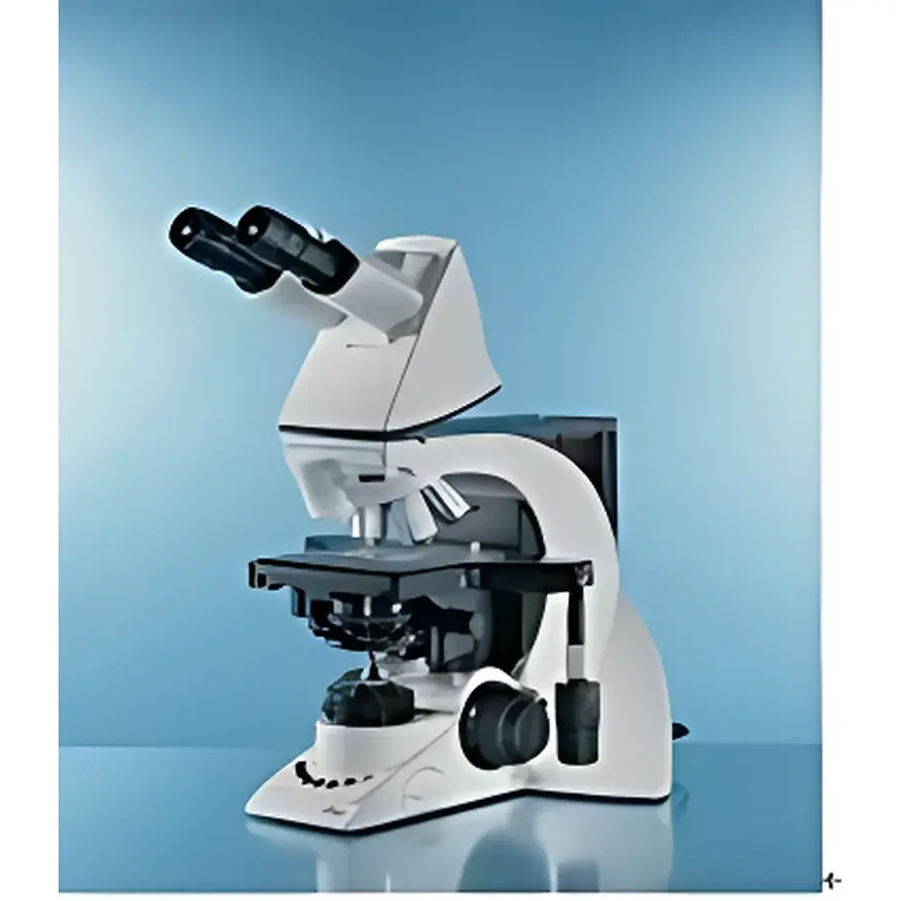
Leica DM3000 Biological Microscope
| Brand | Leica |
|---|---|
| Origin | Germany |
| Model | DM3000 |
| Type | Upright Biological Microscope |
| Illumination | 12 V/30 W Halogen (Transmitted Light) + Optional Fluorescence Light Sources (50 W Hg, 75 W Xe, 100 W Hg) |
| Objective Turret | Motorized 6-Position |
| Condenser | Motorized Achromatic-Aplanatic Flip-Top (1.25×–100×) with Auto-Positioning |
| Illumination Control | Automatic Brightness Adjustment per Objective |
| Focus Mechanism | Height-Adjustable Dual-Speed Coaxial Focus Knobs (Patent DE10340721), Torque-Adjustable, Focus Limit |
| Stage | Ultra-Hard Ceramic XY Stage with Left/Right Hand Reconfigurability, Rotatable Dual-Slide Holder |
| Eyepiece Tube | Ergo Tilting Binocular Tube (15° or 30° inclination, FOV 22 or 25) |
| Contrast Methods | Brightfield (BF), Phase Contrast (PH), Darkfield (DF), Polarization (POL), Differential Interference Contrast (DIC), Fluorescence (FL) |
| Software Compatibility | Leica Application Suite (LAS) for Clinical Imaging & Documentation |
| Compliance | Designed for ISO 13485-aligned workflows |
Overview
The Leica DM3000 Biological Microscope is an upright, modular research-grade instrument engineered for high-throughput clinical and biomedical laboratories. Built on Leica Microsystems’ long-standing optical heritage in German manufacturing, the DM3000 integrates precision mechanical design with intelligent automation to support routine and advanced applications in pathology, cytology, hematology, and basic life science research. Its core optical architecture employs Köhler illumination principles for uniform, glare-free transmitted-light imaging across magnifications from 4× to 100×. The system is inherently compatible with fluorescence modalities through optional high-intensity arc-lamp sources (50 W mercury, 75 W xenon, or 100 W mercury) and a motorized 5-position fluorescence filter changer—enabling rapid spectral selection without manual intervention. Unlike entry-level educational microscopes, the DM3000 features a rigid, thermally stable stand with reinforced optical path alignment, ensuring long-term repeatability and minimal focus drift during extended acquisition sessions.
Key Features
- Motorized 6-position objective turret with patented “Toggle Mode”: enables sub-second switching between two user-defined objectives (e.g., 10× for screening and 40× for diagnostic confirmation), reducing operator fatigue and workflow interruption.
- Auto-positioning achromatic-aplanatic flip-top condenser: automatically moves to the optimal height and aperture setting for each objective magnification (1.25×–100×), maintaining consistent resolution and contrast without manual recalibration.
- Intelligent illumination management: dynamically adjusts halogen lamp intensity based on selected objective—storing preconfigured brightness values per magnification to eliminate manual dimming and reduce photobleaching risk in fluorescence applications.
- Ergonomic focus system: coaxial dual-speed focusing with height-adjustable knobs (patented DE10340721), torque control, and mechanical focus limit—designed for sustained use by users of varying stature and hand size.
- Ultra-hard ceramic XY stage: chemically inert, scratch-resistant surface with symmetrical left/right operability, rotatable dual-slide holder, and smooth, backlash-free movement—even under heavy objective loads.
- Modular tube configuration: interchangeable binocular tubes—including ergo-tilting options (15° or 30° inclination) with field numbers of 22 or 25—allow customization for seated or standing workstations and integration with digital cameras or eyepiece-based measurement accessories.
Sample Compatibility & Compliance
The DM3000 accommodates standard 1″ × 3″ glass slides, Petri dishes (up to 100 mm diameter), and multi-well plates when used with optional stage adapters. Its optical train supports all major contrast techniques required in regulated environments: brightfield for H&E-stained tissue sections; phase contrast for unstained live cells; darkfield for spirochetes or calcifications; polarization for collagen or urate crystals; DIC for subcellular organelle visualization; and fluorescence for immunohistochemistry (IHC), FISH, or GFP-tagged constructs. The microscope’s mechanical stability, repeatable stage positioning, and calibrated magnification scaling meet foundational requirements for ASTM E29-23 (standard practice for using significant digits) and ISO/IEC 17025 traceability frameworks. When paired with Leica Application Suite (LAS) software, the system supports FDA 21 CFR Part 11-compliant electronic records—including user authentication, audit trails, and immutable image metadata logging—making it suitable for GLP and GMP-regulated pathology labs.
Software & Data Management
The DM3000 is fully compatible with Leica Application Suite (LAS) v4.x and later, a CE-marked medical device software platform certified under ISO 13485:2016. LAS provides integrated image capture, annotation, measurement (linear, area, particle count), multi-channel fluorescence overlay, and report generation with customizable templates. Its “Workflow Assistant” module allows technicians to define step-by-step imaging protocols—e.g., auto-focus at 10×, capture overview image, switch to 40×, acquire high-mag field, apply fluorescence channel—reducing inter-operator variability. All acquisitions are time-stamped, user-logged, and stored with EXIF metadata (objective ID, exposure, gain, condenser position). LAS supports DICOM export for PACS integration and HL7-compatible reporting for LIS connectivity—critical for hospital-based cytology and surgical pathology departments.
Applications
The DM3000 serves as a primary diagnostic tool in clinical pathology labs performing frozen section analysis, cervical cytology (Pap smears), bone marrow aspirates, and peripheral blood film review. In academic and pharmaceutical research settings, it supports longitudinal live-cell imaging (with environmental chamber compatibility), routine histology QC, antibody validation via IHC scoring, and training in morphological pattern recognition. Its robust mechanical design and thermal compensation of focus mechanisms ensure reliability during overnight time-lapse experiments. The motorized turret and auto-condenser also accelerate high-volume screening tasks—such as micronucleus assays or comet assays—where rapid objective switching and consistent illumination are essential for statistical validity.
FAQ
Is the DM3000 compliant with FDA 21 CFR Part 11 for electronic records?
Yes—when operated with Leica Application Suite (LAS) configured in audit-trail mode, the system meets electronic signature and record retention requirements for regulated clinical environments.
Can the DM3000 be upgraded to support confocal imaging?
No—the DM3000 is a widefield transmitted-light and epifluorescence platform; confocal capability requires a dedicated Leica TCS SP8 or STELLARIS system.
What fluorescence filter sets are available for the DM3000?
Standard options include DAPI/FITC/TRITC/Cy5 quad-band sets, plus single-band filters for CFP/YFP/mCherry; all mounted in the motorized 5-position filter cube holder.
Does the DM3000 support motorized Z-stack acquisition?
Not natively—the focus drive is motorized but lacks built-in Z-stepping firmware; however, third-party hardware controllers (e.g., Prior ProScan) can be integrated via RS-232 or USB for automated serial sectioning.
Is service and calibration support available outside Germany?
Yes—Leica Microsystems maintains authorized service centers in over 40 countries, with certified field engineers trained in optical alignment, lamp calibration, and mechanical recalibration per ISO 9001 procedures.
Related Products


