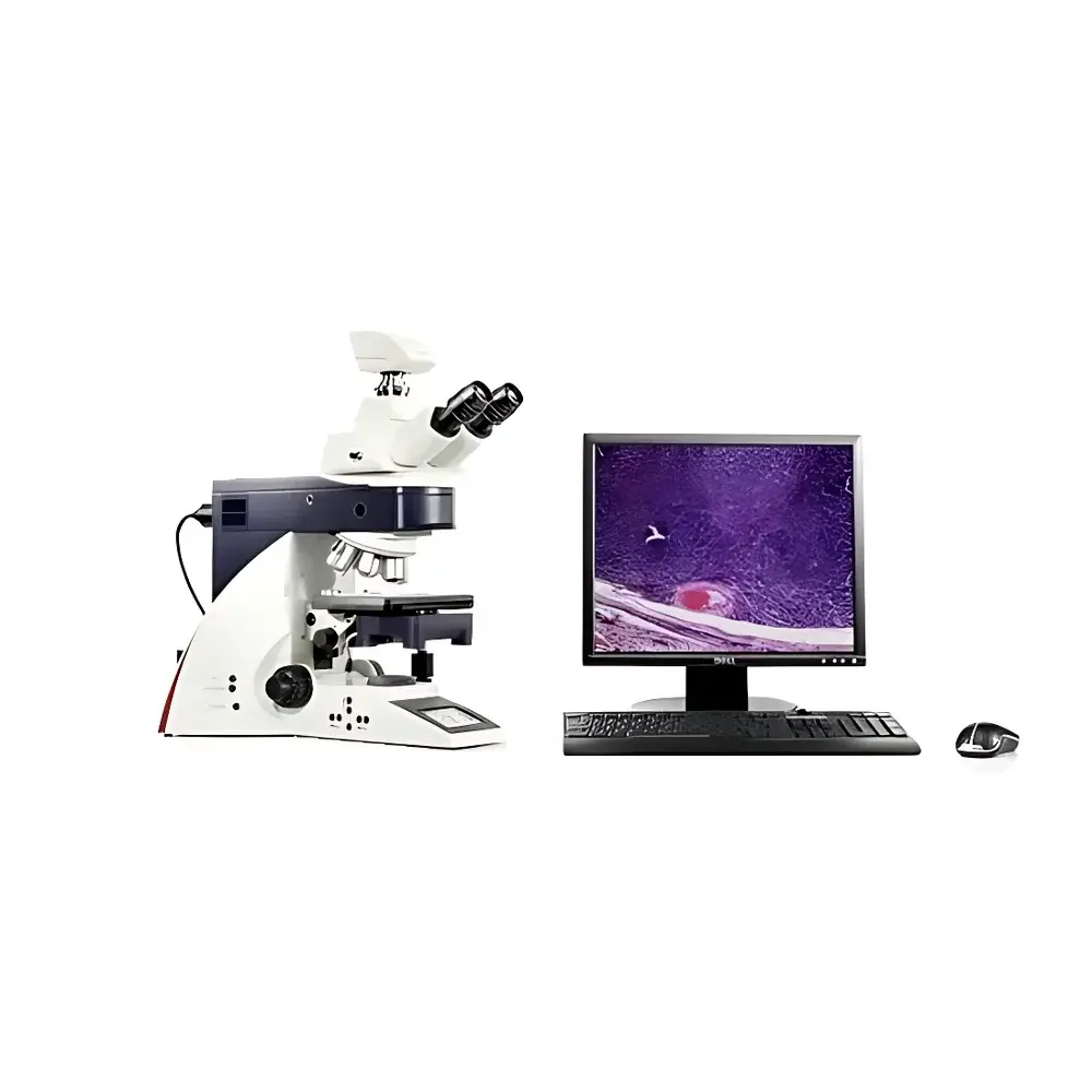
Leica DM4000B Biological Microscope
| Brand | Leica |
|---|---|
| Origin | Germany |
| Model | DM4000B |
| Illumination | Integrated LED transmission and fluorescence illumination |
| Objective Turret | Motorized 6-position |
| Observation Modes | Brightfield, Darkfield, Phase Contrast, Polarization, Fluorescence |
| Software Integration | Compatible with Leica Application Suite (LAS) and third-party imaging platforms |
| Compliance | Designed for GLP/GMP environments |
| Optical Design | Infinity-corrected optical system with Plan Apochromat and Plan Fluotar objective support |
| Eyepiece Field of View | 25 mm |
| Viewing Head | Binocular or trinocular (C-mount port for camera integration) |
| Illumination Control | Automatic intensity tracking, Köhler illumination management, Fluorescence Intensity Management (FIM) technology |
| Fluorescence Module | Motorized 5-position filter cube changer, motorized field diaphragm, shutter, zero-drift filter positioning |
| User Interface | High-resolution OLED parameter display, six programmable ergonomic shortcut keys on focus knobs |
Overview
The Leica DM4000B Biological Microscope is a high-performance, research-grade upright microscope engineered for reproducible, multi-modal imaging in life science laboratories and clinical research settings. Built upon Leica Microsystems’ proven infinity-corrected optical architecture, the DM4000B integrates precision mechanical design with intelligent illumination control to deliver consistent image quality across brightfield, darkfield, phase contrast, polarization, and fluorescence modalities. Its modular platform supports seamless transition between routine histology, live-cell observation, and quantitative fluorescence assays—without manual realignment or recalibration. The system’s core functionality is anchored in its motorized 6-position objective turret, fully automated Köhler illumination management, and patented Fluorescence Intensity Management (FIM) technology, which ensures photostability and inter-session repeatability critical for longitudinal studies and regulatory submissions.
Key Features
- Motorized 6-position objective turret with automatic magnification and correction detection—enables precise, repeatable switching between objectives without user intervention.
- Integrated high-efficiency LED illumination system delivering stable color temperature (5700 K) across full intensity range, with >50,000-hour lifetime and zero warm-up time—eliminating halogen lamp replacement cycles and thermal drift.
- Automatic transmission light intensity tracking: real-time adjustment based on selected objective, condenser setting, and observation mode to maintain optimal signal-to-noise ratio.
- Motorized 5-position fluorescence filter cube changer with zero-drift positioning—ensures pixel-perfect registration across multichannel acquisitions and eliminates mechanical hysteresis.
- Fluorescence Intensity Management (FIM): dynamically regulates excitation intensity per channel to minimize photobleaching while preserving quantitative linearity—essential for comparative expression analysis and time-lapse quantification.
- Six programmable shortcut keys ergonomically positioned on focus knobs—allow one-handed, eyes-on-sample operation for toggling illumination modes, adjusting contrast, capturing images, or launching macros.
- OLED parameter display showing real-time status of objective, magnification, illumination intensity, filter position, condenser aperture, and exposure settings—supports operator verification and SOP compliance.
Sample Compatibility & Compliance
The DM4000B accommodates standard glass slides (1–3 mm thickness), Petri dishes, multi-well plates (up to 96-well format with optional stage inserts), and specialized specimen holders for polarized or DIC applications. Its mechanical stage offers 76 × 52 mm travel range with fine positional repeatability (<1 µm). For regulated environments, the microscope—when operated with Leica LAS X software in validated configuration—supports 21 CFR Part 11 compliance through electronic signatures, audit trails, and user-access-controlled method locking. It meets ISO 13485 design requirements for in vitro diagnostic instrumentation and aligns with ASTM E2877-22 (Standard Guide for Microscopy Image Quality Assessment) and ISO/IEC 17025 documentation expectations for calibration traceability and performance verification.
Software & Data Management
The DM4000B interfaces natively with Leica Application Suite (LAS) X, providing unified control of hardware parameters, acquisition workflows, annotation tools, and metadata embedding (EXIF-compliant TIFF and JPEG2000 export). LAS X supports GLP-compliant experiment logging, including timestamps, operator ID, instrument configuration snapshots, and environmental sensor inputs (when integrated with external humidity/temperature monitors). Third-party compatibility includes Open Microscopy Environment (OME)-compatible APIs, Micro-Manager drivers, and direct integration with MATLAB and Python-based analysis pipelines via Leica’s SDK. All imaging parameters—including exposure time, gain, LED intensity, and filter position—are embedded in image headers for FAIR (Findable, Accessible, Interoperable, Reusable) data stewardship.
Applications
This platform serves as a foundational imaging tool in academic cell biology labs conducting live-cell motility assays, neurobiology labs performing dendritic spine morphology analysis, pathology departments performing routine tissue screening, and pharmaceutical QC units executing GxP-compliant cytotoxicity assays. Its polarization capability supports birefringent crystal identification in toxicology studies, while its phase contrast and darkfield modes enable label-free examination of unstained primary cultures or microorganisms. In developmental biology, the DM4000B—paired with time-lapse modules and environmental chambers—facilitates long-term embryo imaging under controlled CO₂ and temperature conditions. Its robust mechanical stability and vibration-damped base also make it suitable for correlative light-electron microscopy (CLEM) sample pre-screening.
FAQ
Does the DM4000B support DIC (Differential Interference Contrast)?
Yes—when equipped with appropriate Nomarski prisms and strain-free objectives (e.g., Leica Plan Fluotar or HC PL Fluotar series), the DM4000B delivers high-resolution DIC imaging with adjustable shear direction and contrast optimization.
Can I upgrade from halogen to LED illumination post-purchase?
No—the DM4000B LED variant features an integrated, non-interchangeable LED light engine; halogen and LED versions are distinct SKUs with different base assemblies and power supplies.
Is the motorized turret compatible with third-party objectives?
It supports all Leica-branded infinity-corrected objectives with standardized M25 threading; non-Leica objectives may require mechanical adapters and lack automatic magnification recognition or correction collar encoding.
What is the maximum supported camera resolution?
The C-mount port (1× magnification factor) accepts sensors up to 2/3″ format; optimal resolution is achieved with Leica DFC series cameras (e.g., DFC9000 GT, 9.4 MP monochrome), delivering diffraction-limited sampling at 100× oil immersion.
How does FIM ensure quantitative fluorescence reproducibility?
FIM correlates excitation intensity with exposure time and detector gain to maintain constant photon flux per pixel across sessions—verified via NIST-traceable neutral density filters and calibrated photodiode monitoring during system validation.


