Transcend Vivoscope MY780 In Vivo Two-Photon Microscopy System
| Brand | Transcend Vivoscope |
|---|---|
| Model | MY780 |
| Origin | Beijing, China |
| Type | In Vivo Two-Photon Excitation Fluorescence (TPEF) and Second Harmonic Generation (SHG) Microscope |
| Application Domain | Cutaneous Structural & Functional Imaging |
| Regulatory Classification | Research-Use-Only (RUO), CE-compliant optical instrumentation |
Overview
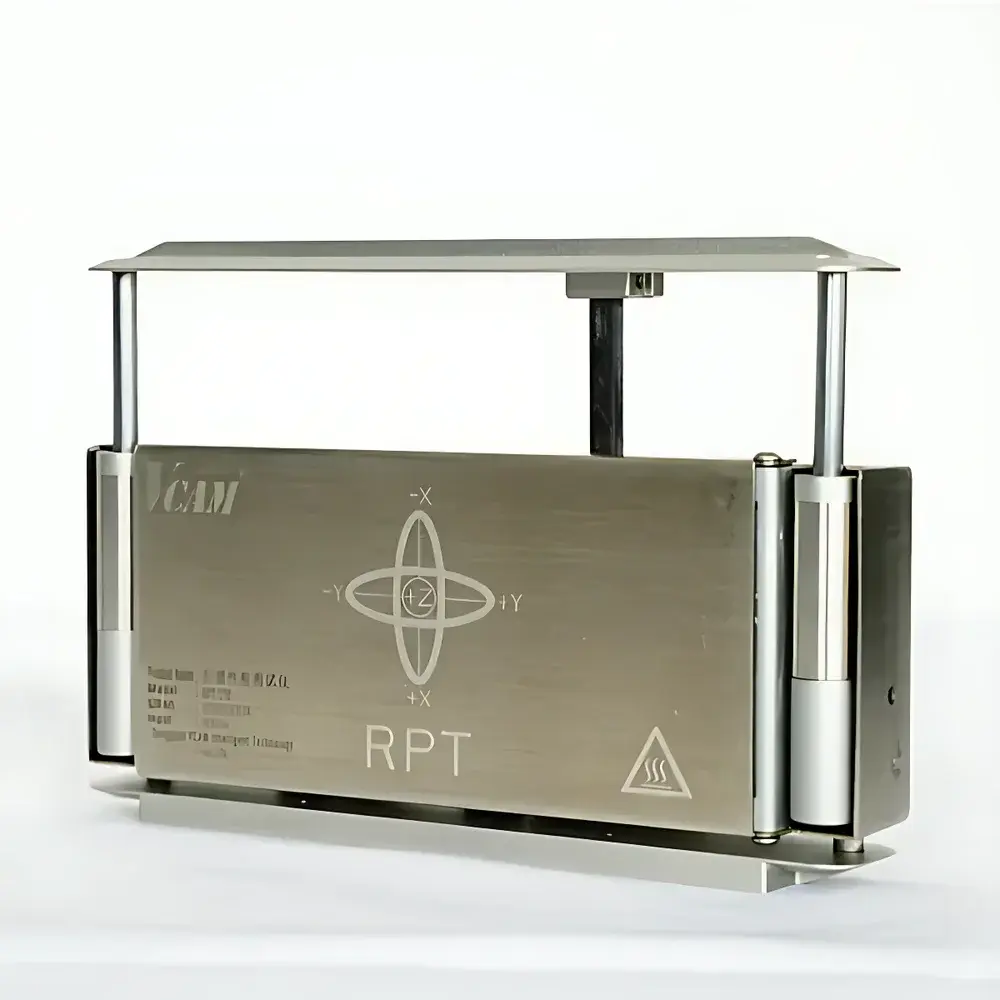
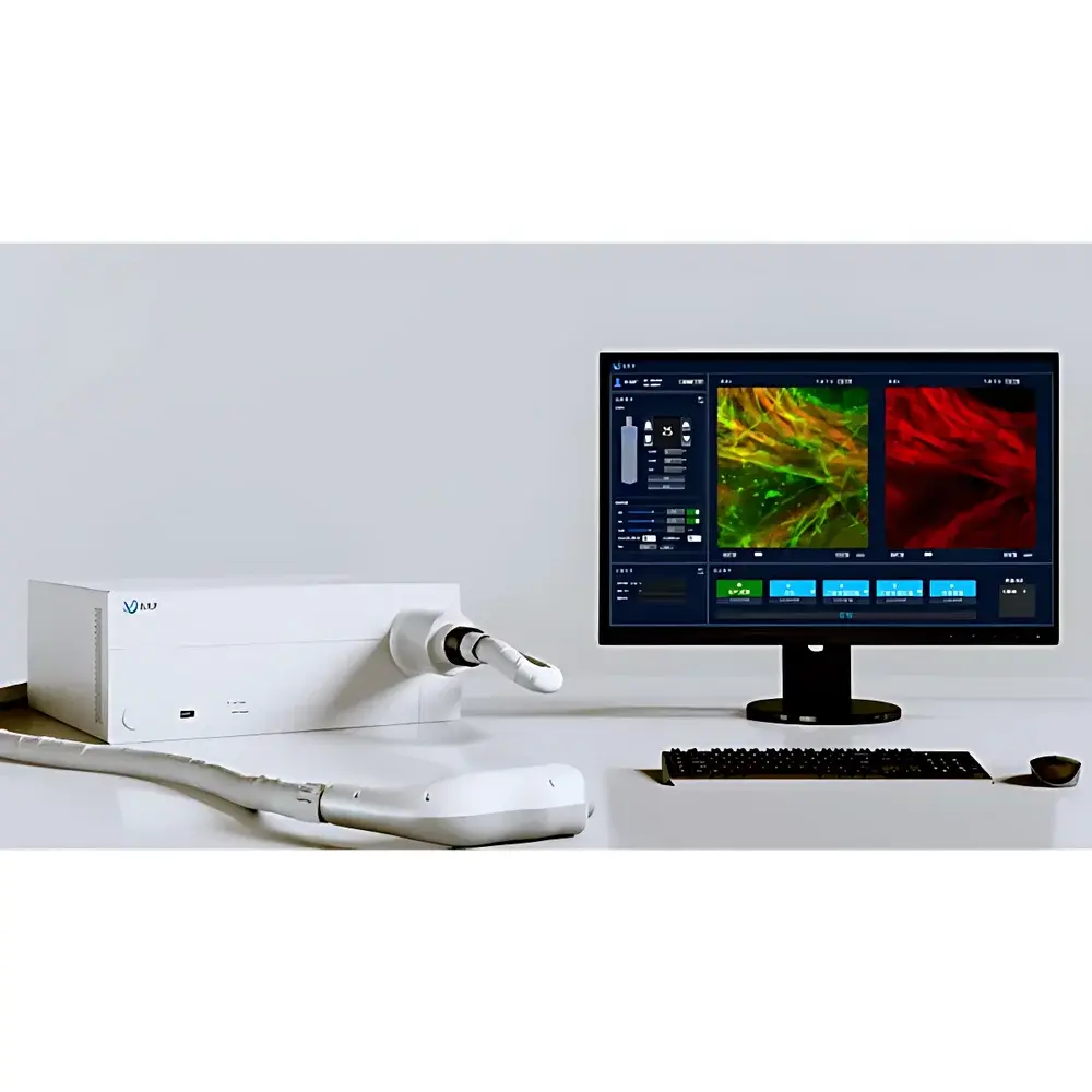
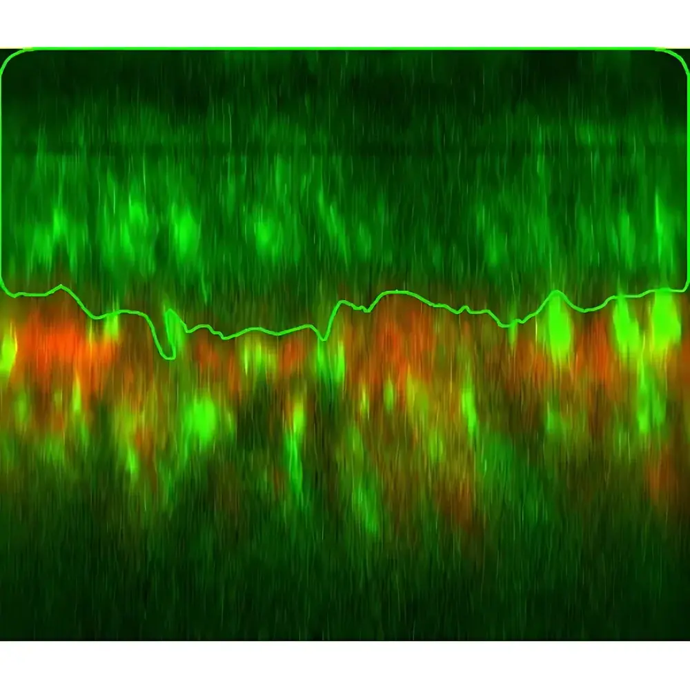
The Transcend Vivoscope MY780 is a compact, turnkey in vivo two-photon microscopy system engineered for high-resolution, label-free, non-invasive imaging of human skin at cellular and subcellular levels. It leverages the physical principles of multiphoton excitation fluorescence (TPEF) and second harmonic generation (SHG) to achieve optical sectioning with inherent depth discrimination—eliminating the need for mechanical sectioning or exogenous contrast agents in many applications. Unlike conventional confocal microscopy, which relies on single-photon excitation and suffers from rapid signal attenuation and phototoxicity in thick tissues, the MY780 employs near-infrared (NIR) pulsed femtosecond lasers (typically 680–1300 nm) to induce simultaneous absorption of two low-energy photons only at the focal plane. This results in highly localized excitation, reduced out-of-focus photobleaching, and significantly improved penetration depth (up to 200–300 µm in human epidermis and upper dermis). SHG imaging further enables structural visualization of endogenous non-centrosymmetric fibrillar proteins—particularly type I and III collagen—without staining, providing quantitative metrics on dermal matrix organization and remodeling.
Key Features
- Integrated dual-modality acquisition: Simultaneous TPEF and SHG channel detection with spectral separation via dichroic mirrors and bandpass filters
- Motorized XYZ scanning stage with sub-micron positioning repeatability for longitudinal in vivo tracking across sessions
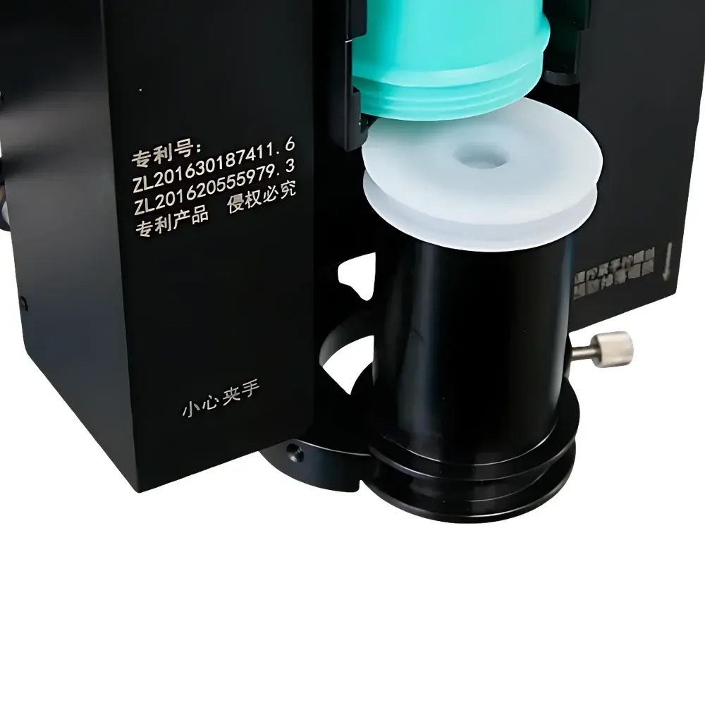
- Dedicated skin interface design: Ergonomic contact-based objective lens housing with adjustable pressure control and real-time epidermal flattening compensation
- Femtosecond Ti:Sapphire laser source (tunable 680–1080 nm) with pulse width <150 fs and repetition rate ≥80 MHz, optimized for minimal thermal load on living tissue
- High-sensitivity GaAsP photomultiplier tubes (PMTs) with analog gain control and photon-counting capability for low-light physiological imaging
- Real-time image preview at ≥15 fps (512 × 512 pixels) and full-resolution acquisition up to 1024 × 1024 pixels with hardware-based descanning and background subtraction
Sample Compatibility & Compliance
The MY780 is validated for use on human volar forearm, cheek, and dorsal hand skin under IRB-approved protocols. It supports both acute imaging sessions (<60 min) and repeated measurements over days/weeks for monitoring dynamic processes such as wound healing, inflammatory infiltration, or pigmentary changes. The system conforms to IEC 60825-1:2014 Class 1M laser safety standards when operated with specified beam delivery optics and skin coupling gel. All optical components meet ISO 10110 surface quality specifications. While designated RUO, its data output structure complies with MIAME-derivative metadata conventions and supports traceable acquisition logs required under GLP-aligned preclinical dermatology studies.
Software & Data Management
Acquisition and analysis are managed through Vivoscope Suite v3.2—a modular, Windows-based platform supporting DICOM-SR export, HDF5-native storage, and batch processing pipelines. Core modules include Z-stack reconstruction with deconvolution (Richardson-Lucy algorithm), SHG/TPEF intensity ratio mapping for collagen-to-cell density quantification, and automated epidermal thickness segmentation using gradient-based edge detection. Audit trails record operator ID, timestamp, laser power, PMT gain, and environmental temperature/humidity per frame. Software architecture supports 21 CFR Part 11-compliant user access control and electronic signature workflows when deployed in regulated environments.
Applications
- Non-invasive assessment of epidermal barrier integrity via keratinocyte morphology and intercellular spacing
- Quantitative dermal collagen architecture analysis in photoaging, scleroderma, and keloid formation
- In vivo tracking of melanosome distribution and transfer dynamics in melasma and vitiligo models
- Real-time vascular perfusion mapping using endogenous NAD(P)H autofluorescence and elastin SHG contrast
- Evaluation of topical drug penetration kinetics by co-localizing fluorescently tagged actives with stratum corneum lipid lamellae
- Longitudinal monitoring of immune cell migration (e.g., Langerhans cell motility) during allergic contact dermatitis induction
FAQ
Is the MY780 suitable for clinical diagnostics?
No—it is classified as Research Use Only (RUO) and not cleared by FDA or CE-IVD for diagnostic decision-making.
Can it image through hair follicles or sebaceous glands?
Yes; its NIR excitation enables partial visualization of infundibular structures and associated pilosebaceous unit activity, though resolution is limited by scattering heterogeneity.
What sample preparation is required?
None beyond standard skin cleansing and application of immersion gel (refractive index matched to stratum corneum); no biopsy, fixation, or staining is needed.
Does it support time-lapse imaging of cellular motility?
Yes; with optional environmental chamber integration (temperature/humidity control), it achieves stable 30-minute+ time-series at 30-second intervals without significant drift.
How is calibration maintained across users and sites?
Each system ships with NIST-traceable reference slides for lateral resolution (1951 USAF target) and intensity linearity verification; software enforces daily dark-current and flat-field correction routines.