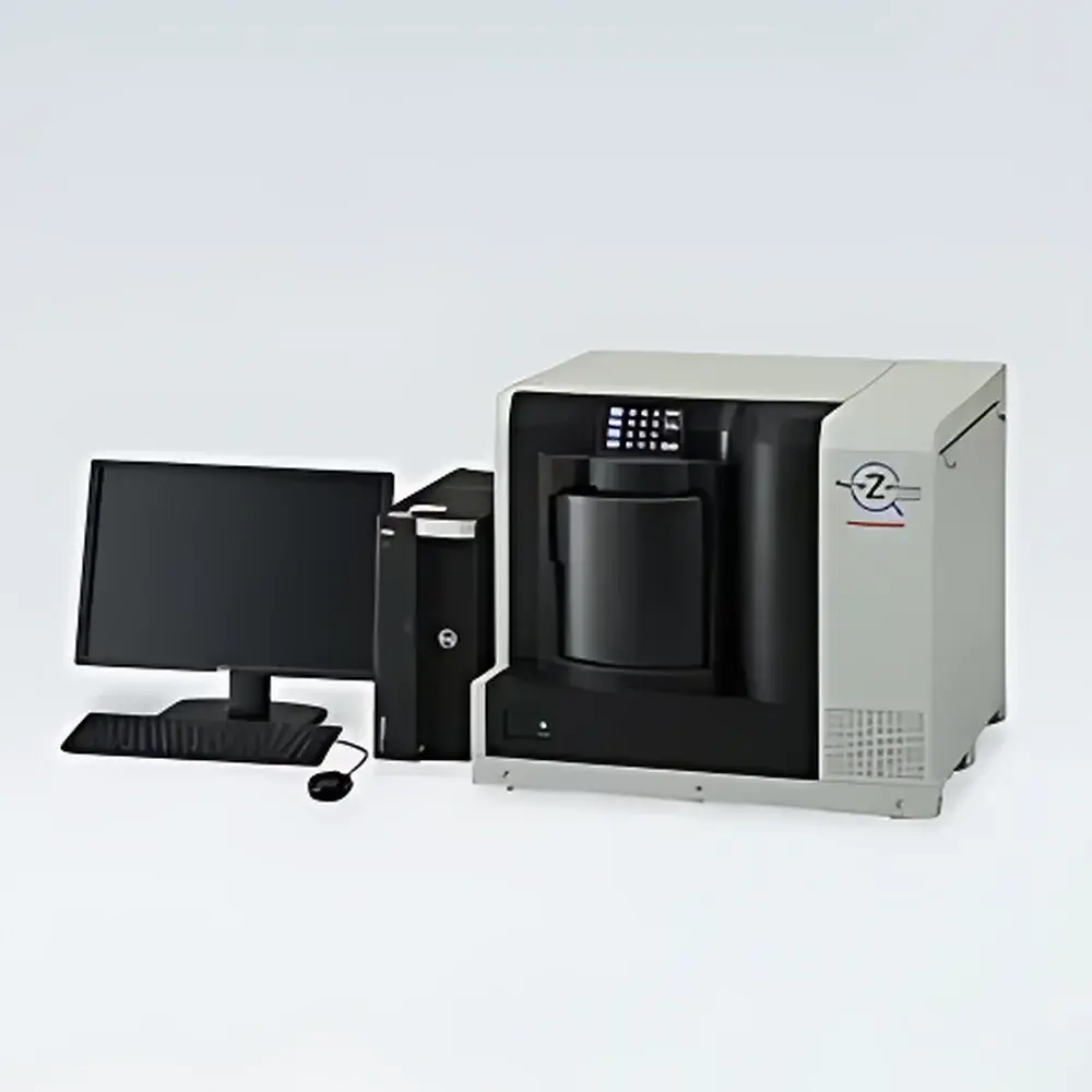
Hamamatsu NanoZoomer S360 Digital Slide Scanner C13220-01
| Brand | Hamamatsu |
|---|---|
| Origin | Japan |
| Manufacturer Type | Original Equipment Manufacturer (OEM) |
| Product Category | Imported Instrument |
| Model | C13220-01 |
| Application Field | Research |
| Imaging Modality | Brightfield Scanning |
| Scan Coverage | Whole-Slide Imaging (WSI) |
| Throughput | High-Throughput |
| Operation Mode | Fully Automated |
| Form Factor | Benchtop |
| Spatial Resolution | 0.23 µm/pixel (40×), 0.46 µm/pixel (20×) |
| Slide Capacity | 360 standard glass slides (26 mm × 76 mm, thickness 0.9–1.2 mm) |
Overview
The Hamamatsu NanoZoomer S360 Digital Slide Scanner (Model C13220-01) is a high-throughput, brightfield whole-slide imaging (WSI) system engineered for precision digitization of histopathological and cytological specimens. Utilizing high-stability motorized stage control, telecentric optical path design, and synchronized line-scan CCD acquisition, the S360 captures gigapixel-resolution digital slides with exceptional geometric fidelity and photometric consistency. Its core architecture supports both 20× (NA 0.75) and 40× objective-based scanning modes—each delivering sub-micron spatial sampling (0.46 µm/pixel at 20×; 0.23 µm/pixel at 40×) without interpolation or pixel binning. Designed for integration into clinical pathology workflows and translational research pipelines, the S360 complies with DICOM Supplement 145 (Whole Slide Imaging) and supports standardized metadata embedding per ISO/IEC 23008-17 (MPEG-H Part 17). The system operates as a Class I medical device in jurisdictions where registered (e.g., China, Taiwan, Japan, Korea, Canada, Turkey, Israel), while designated for research use only (RUO) in regions lacking regulatory clearance for diagnostic applications.
Key Features
- Fully automated benchtop platform with integrated 360-slide capacity loader (30 slides × 12 cassette slots), enabling unattended overnight scanning campaigns.
- Dual-magnification scanning: selectable 20× (0.46 µm/pixel) or 40× (0.23 µm/pixel) objectives with identical 15 mm × 15 mm field-of-view capture time (~30 seconds per FOV), ensuring consistent throughput across magnifications.
- High-throughput performance: >82 slides/hour under both 20× and 40× scanning protocols (measured using 5-focus Z-stack per slide).
- Precision autofocus via pre-scan focus map generation and real-time Z-stack optimization, minimizing out-of-focus artifacts across heterogeneous tissue sections.
- Standard 1D barcode reader (optional 2D support) for automated slide identification and LIMS integration; all barcodes embedded directly into DICOM-SR and JPEG2000 metadata headers.
- Robust thermal management and vibration-damped optical enclosure to maintain focus stability during extended acquisition sessions (>12 hours continuous operation).
Sample Compatibility & Compliance
The NanoZoomer S360 accepts standard microscope slides conforming to ISO 8573-1:2010 dimensions (26 mm × 76 mm) and thickness specifications (0.9–1.2 mm). It supports conventional H&E, IHC, special stains (e.g., Masson’s trichrome, PAS), and cytology preparations on glass substrates. No fluorescent module is included in the base configuration (C13220-01); fluorescence-capable variants require separate validation and hardware integration. Regulatory status varies by jurisdiction: the C13220-01 model is registered as an IVD device in China (NMPA), Taiwan (TFDA), Japan (PMDA), Korea (MFDS), Canada (Health Canada MDEL), Turkey (TİTCK), and Israel (MoH). In the EU and UK, only the CE-marked C13220-21MDEU variant complies with IVDR (EU) 2017/746 and UK MDR 2002. Per EU Commission guidance, the C13220-01 was withdrawn from IVDD (98/79/EC) distribution after 27 May 2022. In the United States, the FDA-cleared C13220-01MD variant is validated for primary diagnosis when paired with FDA-approved displays (e.g., JVC JD-C240BN01A, Barco MDPC-8127) per 21 CFR Part 809.10.
Software & Data Management
The NanoZoomer S360 operates exclusively with Hamamatsu’s NDP.scan software v3.x or later, which provides DICOM-compliant image export (JPEG2000 compression with lossless/lossy options), multi-layer pyramid tiling (OpenSlide-compatible), and embedded annotation support. All acquired data includes EXIF and DICOM-SR metadata fields covering scanner ID, objective, exposure parameters, focus map coordinates, and barcode identifiers. Audit trails meet GLP/GMP requirements: software logs record user login/logout, scan start/stop timestamps, parameter modifications, and error events with immutable timestamps. For enterprise deployment, optional NDP.view Enterprise Server enables role-based access control, HL7/DICOM modality worklist integration, and PACS interfacing via standard DICOM WADO-URI and QIDO-RS APIs. Data integrity is enforced through SHA-256 hash verification upon ingestion and storage.
Applications
- Clinical pathology departments requiring high-volume digitization for telepathology consultation, second-opinion review, and archival compliance (e.g., CAP accreditation requirements for digital storage).
- Academic and pharmaceutical research laboratories conducting quantitative image analysis (QIA) of tumor-infiltrating lymphocytes (TILs), mitotic counts, or spatial biomarker mapping using AI-driven segmentation tools.
- Biobank operations performing longitudinal slide re-scanning for quality assurance, stain normalization studies, or retrospective cohort digitization projects.
- Regulatory submission support: generation of audit-ready digital slide sets compliant with FDA eCTD Module 5.3.5.2 (digital pathology evidence) and EMA CHMP/ICH guidelines on analytical validation of computational pathology algorithms.
- Education and training: creation of annotated virtual slide libraries for pathology residency programs aligned with ACGME Milestones and Royal College of Pathologists curriculum standards.
FAQ
Is the C13220-01 approved for primary diagnosis in the United States?
Yes—only the FDA-cleared C13220-01MD variant (not C13220-01) is authorized for primary diagnostic use in the U.S., contingent upon use with FDA-approved display monitors and adherence to 21 CFR Part 11 electronic signature requirements.
Does the NanoZoomer S360 support fluorescence scanning?
No—the C13220-01 configuration is optimized exclusively for brightfield imaging. Fluorescence-capable models (e.g., NanoZoomer S60 or S540 with optional filter cubes) require separate regulatory validation and are not interchangeable with the S360 platform.
What DICOM conformance profiles does the S360 implement?
The system conforms to DICOM Supplement 145 (Whole Slide Imaging), including WSI Storage SOP Class (1.2.840.10008.5.1.4.1.1.77.1.6), Basic Text SR (1.2.840.10008.5.1.4.1.1.88.11), and Modality Worklist (1.2.840.10008.5.1.4.1.1.2).
Can the S360 be integrated into existing LIS/PACS environments?
Yes—via native DICOM interfaces (WADO-URI, QIDO-RS, STOW-RS) and optional HL7 v2.x messaging adapters provided through Hamamatsu’s NDP.view Enterprise Server licensing tier.
What is the expected service life and maintenance schedule?
Hamamatsu recommends biannual preventive maintenance (PM) visits by certified field service engineers, including optical alignment verification, stage calibration, and CCD sensor dark-frame characterization. Mean time between failures (MTBF) exceeds 15,000 operational hours under standard laboratory conditions (23°C ± 2°C, 40–60% RH).


