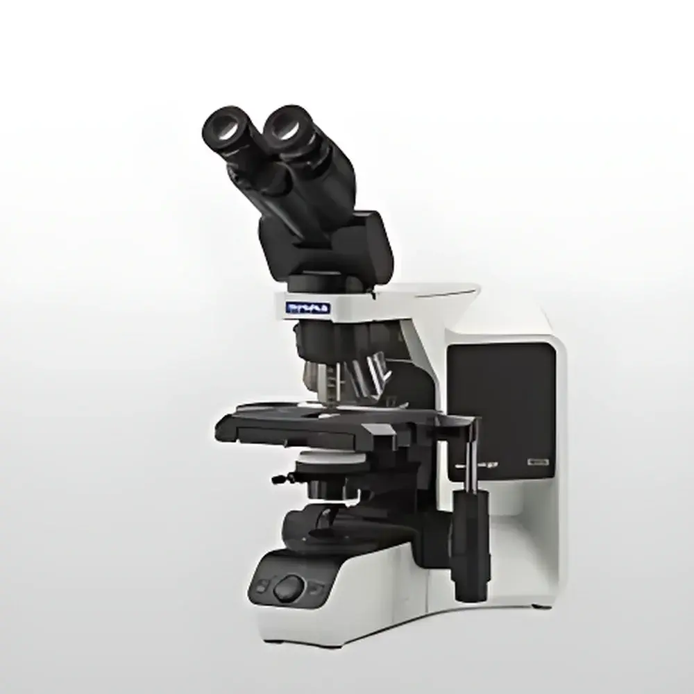
Olympus BX43 Upright Biological Microscope
| Brand | Olympus |
|---|---|
| Origin | Japan |
| Manufacturer | Olympus Corporation |
| Product Type | Upright Microscope |
| Model | BX43 |
| Light Source | High-CRI White LED (equivalent to 30 W halogen, stable color temperature across intensity levels, 20,000 h lifetime) |
| Ergonomic Design | Adjustable observation tube and mechanical stage |
| Modularity | Fully modular optical platform for configuration scalability |
Overview
The Olympus BX43 is a high-performance upright biological microscope engineered for routine and advanced life science applications in academic laboratories, clinical pathology units, and industrial quality control environments. Built upon Olympus’ proven UIS2 optical system, the BX43 delivers exceptional image fidelity, flat-field correction, and chromatic aberration control across wide-field eyepieces and digital imaging paths. Its core design follows the principles of Köhler illumination and infinity-corrected optics, ensuring consistent contrast, resolution, and color fidelity regardless of magnification or accessory configuration. As a modular platform, the BX43 supports seamless integration of phase contrast, differential interference contrast (DIC), fluorescence, and brightfield modalities—enabling researchers to adapt the system precisely to evolving experimental requirements without hardware redundancy or workflow disruption.
Key Features
- Modular Optical Architecture: Interchangeable observation tubes (binocular, trinocular, inclined/rotatable), nosepieces (5×–6× objective turrets), and condensers (Abbe, swing-out, centerable) allow configuration optimization for teaching, diagnostics, or multi-modal imaging.
- High-Color-Fidelity LED Illumination: A 3W white LED light source provides uniform, flicker-free illumination with a correlated color temperature of 5,700 K across all intensity settings—matching daylight-balanced microscopy standards without thermal drift or bulb replacement cycles.
- Ergonomic Workflow Integration: Low-positioned coaxial focus controls, extended-stage mechanical controls, and an adjustable-height observation tube reduce operator fatigue during prolonged use—aligned with ISO 9241-5 ergonomic guidelines for visual display workstations.
- UIS2 Optical Platform: All objectives (including Plan N, UPLSAPO, and LCAch series) are optimized for the 200 mm tube length and 22 mm field number, delivering diffraction-limited resolution at 100× oil immersion (NA 1.40) and minimal spherical aberration under live-cell conditions.
- Robust Mechanical Stability: The rigid cast-aluminum frame, anti-vibration base, and precision-ground stage rails ensure positional repeatability and vibration damping—critical for time-lapse imaging and quantitative morphometric analysis.
Sample Compatibility & Compliance
The BX43 accommodates standard 1 mm-thick glass slides (76 × 26 mm), petri dishes (up to 100 mm diameter), and culture flasks via optional stage adapters. It supports live-cell observation using heated stage inserts (20–45 °C range) and CO2-compatible environmental chambers. All optical components comply with ISO 8578 (microscope nomenclature), ISO 10934-1 (optical resolution testing), and JIS B 7151 (Japanese Industrial Standard for microscopes). For regulated environments, the system supports GLP-compliant documentation when paired with Olympus cellSens software featuring 21 CFR Part 11–ready audit trails, electronic signatures, and user-access-level management.
Software & Data Management
When integrated with Olympus cellSens imaging software (v3.0+), the BX43 enables synchronized hardware control, multi-channel fluorescence acquisition, Z-stack reconstruction, and quantitative intensity profiling. The software architecture supports DIC vector analysis, particle counting with size-classified thresholds, and time-series registration for motility studies. Data export conforms to TIFF 6.0, OME-TIFF, and JPEG2000 standards—ensuring interoperability with ImageJ/Fiji, MATLAB, and commercial LIMS platforms. Raw metadata (objective NA, magnification, exposure time, LED intensity, stage coordinates) is embedded per frame in EXIF-compatible headers.
Applications
- Routine histopathology screening and cytology assessment in clinical laboratories
- Live-cell imaging of adherent and suspension cultures under controlled thermal and gaseous conditions
- Developmental biology studies requiring long-term timelapse of embryonic tissues
- Microbiology workflows including Gram staining evaluation and fungal morphology analysis
- Pharmaceutical QC for particulate matter inspection in parenteral preparations (per USP )
- Academic teaching labs where modularity supports progressive skill development—from basic brightfield to advanced contrast techniques
FAQ
Is the BX43 compatible with third-party digital cameras?
Yes—the trinocular port features a standardized C-mount interface (1× or 0.5× photo factor options) and supports USB 3.0 and HDMI output via Olympus DP-series or industry-standard CMOS sensors.
Can the BX43 be upgraded for fluorescence imaging?
Yes—fluorescence capability is added via filter cube sliders (U-MF2, U-MFPH), mercury or LED excitation sources (e.g., U-HGLGPS), and high-NA objectives; no optical realignment is required.
What maintenance is required for the LED illumination system?
None beyond periodic cleaning of the collector lens and dust cover; the LED driver includes thermal regulation and current stabilization to maintain photometric consistency over its rated 20,000-hour service life.
Does Olympus provide validation documentation for GMP environments?
Yes—Olympus offers IQ/OQ protocols, calibration certificates traceable to NIST standards, and software validation packages aligned with Annex 11 and FDA guidance for computerized systems.
Related Products


