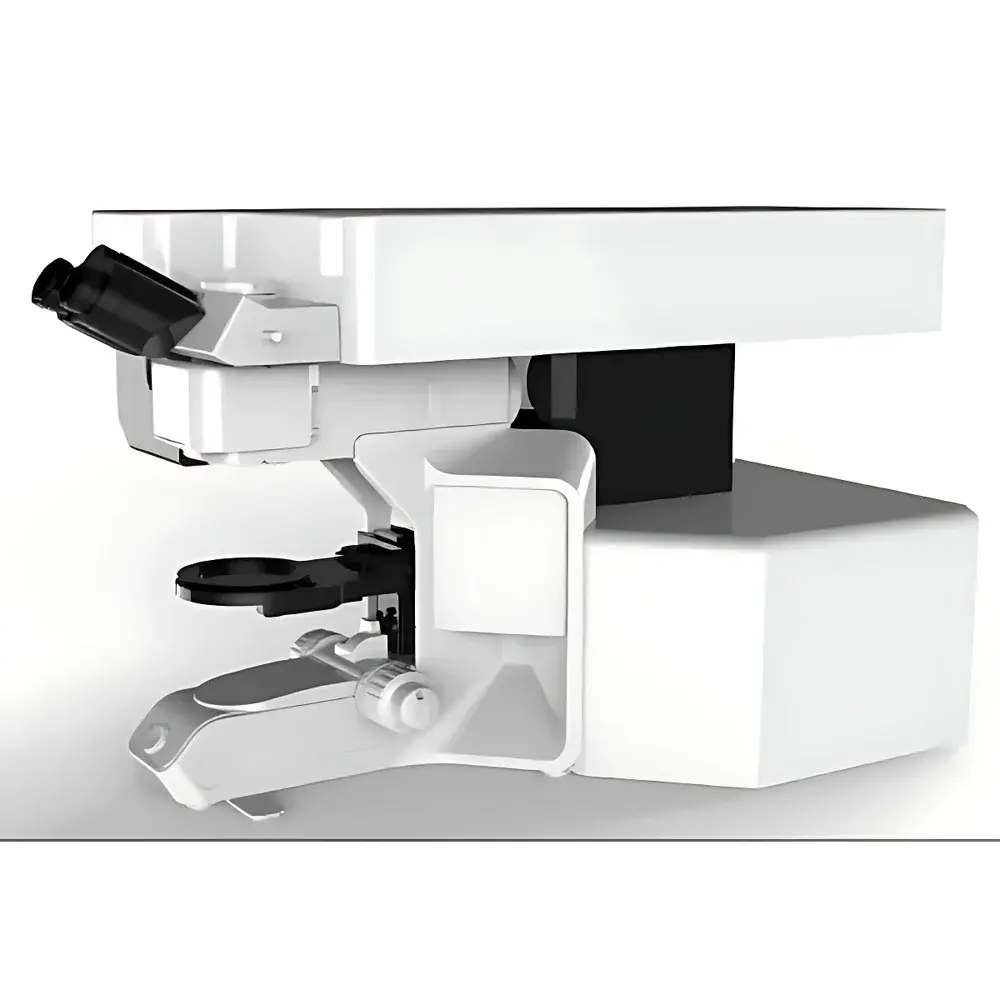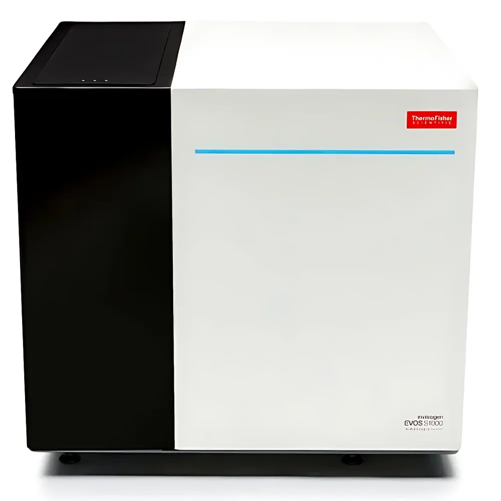
Invitrogen EVOS S1000 Spatial Biology Imaging and Analysis System
| Brand | Invitrogen |
|---|---|
| Origin | USA |
| Manufacturer Type | Original Equipment Manufacturer (OEM) |
| Import Status | Imported |
| Model | EVOS S1000 |
| Pricing | Available Upon Request |
| Sensor Resolution | 4.2 Megapixels |
| Camera Type | 16-bit sCMOS |
| Objective Turret | 5-position, supporting 2.5× to 40× objectives |
Overview
The Invitrogen EVOS S1000 Spatial Biology Imaging and Analysis System is a benchtop, integrated fluorescence and brightfield imaging platform engineered for high-fidelity spatial profiling of tissue sections at cellular and subcellular resolution. Built upon a robust optical architecture incorporating precision motorized stage control, multi-channel LED excitation, and a thermally stabilized 16-bit scientific CMOS (sCMOS) sensor, the system enables quantitative, reproducible acquisition of multiplexed spatial data without requiring confocal scanning or complex alignment procedures. Its core design adheres to principles of widefield epifluorescence microscopy with hardware-optimized spectral separation and low-noise detection—making it suitable for both discovery-phase spatial omics validation and translational pathology workflows. The EVOS S1000 operates as a closed-system imaging solution, minimizing photobleaching through intensity-controlled illumination and delivering consistent signal-to-noise ratios across large-area acquisitions (up to 1 cm² in under 60 minutes at 20× magnification).
Key Features
- High-throughput spatial imaging: Completes full-tissue, 9-plex fluorescent image acquisition over 1 cm² area in ≤60 minutes at 20× objective magnification.
- Multi-modal imaging capability: Simultaneous support for multiplex fluorescence, transmitted brightfield, phase contrast, and color brightfield imaging—enabling correlative morphological and molecular analysis on the same slide.
- Optimized detection sensitivity: 4.2-megapixel sCMOS sensor with 16-bit dynamic range ensures accurate quantification of low-abundance epitopes and fine-grained spatial patterns in complex tissue architectures.
- Flexible optical configuration: 5-position motorized objective turret accommodates standard high-NA objectives ranging from 2.5× to 40×, allowing seamless scaling between whole-slide overview and single-cell resolution.
- Automated acquisition workflow: Integrated autofocus, auto-exposure, and tile-based stitching eliminate manual intervention—reducing operator variability and improving inter-run reproducibility.
- Intuitive spatial analysis interface: EVOS™ Spatial Software provides guided, modular analysis pipelines—from registration and spectral unmixing to cell segmentation, neighborhood analysis, and spatial clustering—designed for users with minimal computational training.
Sample Compatibility & Compliance
The EVOS S1000 accepts standard 1 × 3 inch glass microscope slides, including FFPE and frozen tissue sections up to 10 µm thickness, as well as cytospin preparations and cultured monolayers. It supports common spatial proteomics and transcriptomics assay formats, including antibody-conjugated detection reagents (e.g., tyramide signal amplification, polymer-based secondary systems) and commercially available oligonucleotide-tagged probes. All imaging protocols are compatible with routine histopathology workflows and validated against ASTM E2877-21 (Standard Guide for Digital Image Acquisition in Microscopy) and ISO/IEC 17025:2017 requirements for measurement traceability. Data integrity is maintained through embedded audit trails, user authentication, and timestamped acquisition logs—supporting GLP-compliant documentation and FDA 21 CFR Part 11 readiness when deployed in regulated environments.
Software & Data Management
EVOS™ Spatial Software serves as the unified acquisition and analysis engine, featuring built-in calibration tools, batch processing queues, and export options compliant with OME-TIFF and Bio-Formats standards. The software includes an onboard dye library with pre-validated excitation/emission profiles for >30 commonly used fluorophores—including Alexa Fluor®, DyLight®, and Cy® dyes—as well as compatibility with custom spectral signatures for novel conjugates. Quality metrics (e.g., focus score, signal uniformity, channel crosstalk index) are computed in real time and compiled into automated QC reports. Raw and processed datasets are structured using MIAME-compliant metadata schemas and support integration with downstream spatial analytics platforms such as Visium, Xenium, or open-source tools like Squidpy and Seurat via standardized HDF5 or .csv exports.
Applications
- Tumor microenvironment characterization: Maps immune cell infiltration, stromal interactions, and spatially resolved biomarker co-expression to identify predictive signatures for immunotherapy response.
- Neuroscience research: Resolves laminar organization, synaptic protein distribution, and glial-neuronal spatial relationships in human postmortem and rodent brain sections.
- Drug target validation: Quantifies on-target engagement, pathway modulation, and off-target binding patterns across tissue compartments in preclinical efficacy and toxicity studies.
- Clinical assay development: Supports analytical validation of spatial biomarkers for companion diagnostic use, including reproducibility assessment across laboratories and instrument platforms.
FAQ
What sample preparation methods are compatible with the EVOS S1000?
Standard IHC, IF, H&E, and special stains on deparaffinized FFPE or cryosections are fully supported. Antigen retrieval and blocking steps follow established protocols; no specialized mounting media or coverslipping is required.
Does the system support spectral unmixing for highly overlapping fluorophores?
Yes—EVOS™ Spatial Software includes linear unmixing algorithms calibrated using reference spectra acquired from single-stained control slides.
Can acquired images be exported for third-party analysis?
All images are saved in OME-TIFF format with embedded metadata, enabling direct import into ImageJ/Fiji, QuPath, HALO, or Python-based spatial analysis frameworks.
Is remote operation or multi-user access supported?
The system supports network-enabled acquisition control and centralized data storage via SMB/CIFS shares; role-based user permissions are configurable within the software.
How is system performance verified over time?
Built-in daily calibration routines assess LED intensity stability, stage positioning accuracy, and camera dark current—generating pass/fail logs aligned with ISO 10993-12 verification guidelines.