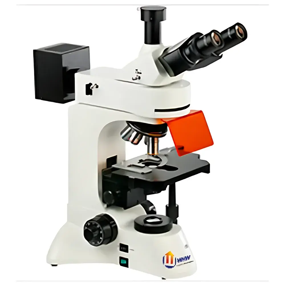
YANRUN FM-600 Upright Infinity-Corrected LED Epi-Fluorescence Microscope
| Brand | YANRUN |
|---|---|
| Origin | Shanghai, China |
| Manufacturer Type | Direct Manufacturer |
| Region | Domestic (China) |
| Model | FM-600 Upright Infinity-Corrected LED Epi-Fluorescence Microscope |
| Price | USD 1 (Base Configuration Only) |
Overview
The YANRUN FM-600 is an upright, infinity-corrected epi-fluorescence microscope engineered for routine and advanced life science imaging applications in academic laboratories, clinical pathology units, and industrial quality control environments. It employs a Köhler-illuminated epi-fluorescence optical path with modular LED excitation sources—eliminating mercury lamp hazards, thermal drift, and spectral instability while delivering stable, narrow-band excitation across UV, violet, blue, and green spectral regions. The system integrates a high-NA Abbe condenser for transmitted-light brightfield and phase contrast observation, and supports simultaneous dual-channel fluorescence acquisition via interchangeable filter sets. Designed around an ergonomic 30° inclined trinocular head with parfocal, wide-field WF10X/22mm eyepieces, the FM-600 ensures operator comfort during extended microscopy sessions and seamless integration with digital imaging systems.
Key Features
- Infinity-corrected optical architecture with plan achromat objectives (4X/0.10, 10X/0.25, 40X/0.65 spring-loaded, 100X/1.25 oil immersion), ensuring minimal chromatic aberration and consistent resolution across magnifications from 40× to 1000×.
- LED-based epi-fluorescence illumination system featuring four independently switchable excitation bands: UV (320–380 nm), Violet (380–415 nm), Blue (410–490 nm), and Green (475–550 nm), each paired with matched emission filters (e.g., 515 nm for B, 595 nm for G).
- High-stability, low-heat 3W white LED transmitted-light source with continuously adjustable intensity and integrated field diaphragm; coupled with an NA 1.25 Abbe condenser with coaxial height adjustment.
- Five-position inward-facing nosepiece with precision ball-bearing positioning and parfocal alignment; coarse/fine coaxial focusing mechanism with 2 µm fine-focus graduation, mechanical stop, and locking function.
- Large 210 × 140 mm mechanical stage with 75 × 50 mm travel range, compatible with standard specimen slides and multi-well plates; optional graduated wide-field eyepieces and 60X plan achromat objective available.
Sample Compatibility & Compliance
The FM-600 accommodates standard 1″ × 3″ glass slides, coverslips (0.13–0.17 mm thickness), and Petri dishes up to 100 mm diameter. Its optical design complies with ISO 8578:2017 (Microscopes — Requirements for performance and testing) and supports GLP-compliant documentation workflows when paired with FDA 21 CFR Part 11–enabled imaging software (via optional USB/VIDEO interface). Fluorescence filter sets meet ANSI Z80.10 standards for spectral bandwidth fidelity. All LED drivers conform to IEC 61000-6-3 EMC emission limits and IEC 62471 photobiological safety classification (Risk Group 1 for all visible bands; RG2 for UV configuration when used with appropriate shielding).
Software & Data Management
The microscope interfaces seamlessly with third-party imaging platforms via C-mount (1×, 0.5×, 0.4× adapters available) and standardized USB 2.0/VIDEO output. Optional calibration-certified CCD or CMOS cameras support time-lapse acquisition, Z-stack reconstruction, and multi-channel overlay analysis. Image metadata—including objective ID, exposure time, LED channel selection, and stage coordinates—is embedded in TIFF/OME-TIFF formats. When deployed with validated image analysis suites (e.g., ImageJ/Fiji, NIS-Elements, or HALO), the FM-600 supports audit-trail generation, user-access controls, and electronic signature capture—fulfilling core requirements of ISO/IEC 17025 and CLIA-aligned laboratory information management systems.
Applications
- Routine histopathology screening using DAPI, FITC, TRITC, and Cy3/Cy5 labeling protocols.
- Live-cell imaging with low-phototoxicity LED excitation, particularly in GFP/RFP-expressing models under controlled environmental chambers.
- Material science inspection of fluorescently tagged polymers, nanocomposites, and semiconductor thin films.
- Forensic fiber analysis and mineral identification via fluorescence response under UV/V excitation.
- Teaching laboratory use for comparative morphology studies across plant, animal, and microbial specimens.
FAQ
Does the FM-600 support oil immersion imaging at 100× magnification?
Yes—the included PL100X/1.25 oil immersion objective is fully compatible with standard Type A cedarwood oil (n = 1.518) and delivers diffraction-limited resolution at 1000× total magnification when used with the 10X wide-field eyepiece.
Can UV fluorescence be used safely without additional enclosures?
UV excitation (320–380 nm) requires adherence to IEC 62471 guidelines: operators must wear UV-blocking safety goggles and avoid direct exposure to unfiltered beam paths. Optional interlocked UV shutter modules are available for enhanced lab safety compliance.
Is the LED light source replaceable in the field?
All LED modules—including excitation and transmitted-light units—are user-replaceable via standardized M3 mounting and JST-XH connectors, with no optical realignment required post-installation.
What digital camera interfaces are supported?
Standard C-mount (1″ sensor format) is provided; optional 0.4×, 0.5×, and 1× reduction lenses enable optimal pixel matching for sensors ranging from 1/3″ to 4/3″ format. USB 2.0 and analog VIDEO outputs allow compatibility with both modern digital capture systems and legacy recording equipment.
Are calibration certificates traceable to national standards available?
Yes—NIST-traceable magnification verification reports and LED spectral irradiance calibrations (per CIE S 026/E:2019) can be supplied upon request for IQ/OQ validation packages.


