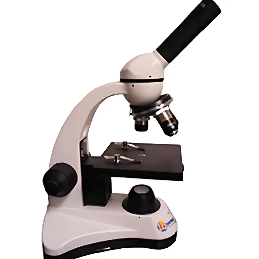
YANRUN BI-11 Biological Inverted Microscope
| Brand | YANRUN |
|---|---|
| Origin | Shanghai, China |
| Manufacturer Type | Direct Manufacturer |
| Product Category | Domestic (China-made) |
| Model | BI-11 Biological Inverted Microscope |
| Instrument Type | Inverted Microscope |
| Magnification Range (Standard) | 40×–400× |
| Max Optional Magnification | 64×–1600× |
| Eyepiece | Wide-Field WF10× (Φ18 mm) |
| Objective Lenses (BI-11A) | 4×/0.10, 10×/0.25, 40×/0.65 (spring-loaded) |
| Objective Lenses (BI-11B) | 4×/0.10, 10×/0.25, 40×/0.65 (spring-loaded), 100×/1.25 (oil immersion, spring-loaded) |
| Eyepiece Tube | Monocular, 360° rotatable, inclined at 45° |
| Focus Mechanism | Coaxial coarse/fine focus, fine focus graduation: 4 µm |
| Nosepiece | BI-11A – 3-position forward-facing ball-bearing turret |
| Stage | BI-11A – fixed stage (120 mm × 120 mm) with 5-hole aperture diaphragm disc |
| Condenser | Abbe condenser (N.A. 1.25), vertically adjustable (BI-11B only) |
| Illumination | High-intensity white LED (integrated or external power supply options), compatible with halogen, tungsten-halogen, fluorescent, and incandescent sources via optional condensers |
| Optional Accessories | WF16× eyepiece, pointer eyepiece, variable-aperture condenser, movable-stage adapter, color filters (green/yellow/blue), ground glass diffuser, USB/VIDEO phototube, rechargeable LED module, 12 V/20 W halogen lamp, 5 W fluorescent lamp, 20 W incandescent lamp |
Overview
The YANRUN BI-11 Biological Inverted Microscope is engineered for routine and advanced life science applications requiring stable, high-contrast observation of living cells, adherent cultures, and semi-transparent specimens in Petri dishes, flasks, and multi-well plates. Unlike upright configurations, its inverted optical architecture positions the objective lenses beneath the specimen stage and the illumination system above—enabling unobstructed access to culture vessels during real-time imaging, micromanipulation, or long-term time-lapse experiments. The system employs standard Köhler illumination principles, with parfocal, achromatic objectives and wide-field eyepieces optimized for ergonomic viewing and consistent image fidelity across magnifications from 40× to 1600× (with oil immersion). Designed for laboratory environments operating under GLP-aligned workflows, the BI-11 complies with IEC 61000-6-3 (EMC emission standards) and meets mechanical safety requirements per ISO 13857 for operator protection during extended use.
Key Features
- Coaxial coarse/fine focusing mechanism with 4 µm fine-focus graduation—enabling precise Z-axis positioning essential for serial sectioning and focal plane tracking;
- Forward-facing ball-bearing nosepiece (3-position standard, 4-position optional) ensuring repeatable, low-friction objective alignment and long-term rotational stability;
- High-brightness white LED illumination (6,500 K CCT, >10,000 h lifetime) with uniform intensity distribution and minimal thermal load on live samples;
- Dual-stage configuration: fixed stage (BI-11A) for static slide-based work; double-layer mechanical stage (BI-11B) with 70 mm × 30 mm travel range for coordinated sample navigation during multi-location imaging;
- Abbe condenser (N.A. 1.25, vertically adjustable) included with BI-11B variant—supporting critical aperture control for optimal resolution and contrast in brightfield and phase contrast modalities;
- Monocular tube with 45° inclination and full 360° rotation—facilitating flexible ergonomics and integration with auxiliary imaging modules;
- Modular illumination compatibility: interchangeable condensers support LED, halogen (12 V/20 W), fluorescent (5 W), and incandescent (20 W) light sources per application-specific requirements.
Sample Compatibility & Compliance
The BI-11 accommodates standard tissue culture formats including 35 mm, 60 mm, and 100 mm Petri dishes; T-25, T-75, and T-175 flasks; and 6-, 12-, 24-, 48-, and 96-well plates—without requiring coverslip mounting. Its stage clearance (≥45 mm) and objective working distances (e.g., 17.5 mm for 10×, 0.65 mm for 40×, 0.13 mm for 100× oil) ensure compatibility with thick-bottomed vessels and gel-based assays. All optical components meet ISO 8578 (microscope performance testing) and JIS B 7151 (achromatic objective specifications). The instrument supports documentation traceability in regulated environments: when paired with optional USB/VIDEO phototube and compliant acquisition software, it can be configured to satisfy audit trail and electronic signature requirements aligned with FDA 21 CFR Part 11 Annex 11 principles.
Software & Data Management
While the BI-11 operates as a standalone optical platform, its integrated USB/VIDEO phototube output (optional) enables seamless connection to third-party image acquisition systems—including open-source platforms like Micro-Manager and commercial suites such as NIS-Elements or ZEN Blue. Video streaming is supported at up to 30 fps (720p) with minimal latency. Metadata tagging (magnification, objective ID, exposure time, timestamp) is preserved in TIFF and AVI export formats. For laboratories implementing digital pathology or QC workflows, the microscope integrates into existing LIMS or ELN infrastructure via standardized DICOM-SR or CSV metadata injection protocols. Firmware updates (where applicable) are delivered via secure HTTPS endpoint with SHA-256 signature verification.
Applications
- Cell culture monitoring: proliferation assessment, confluence estimation, and morphological screening in primary and immortalized lines;
- In vitro toxicology assays: real-time observation of cytotoxicity endpoints (membrane integrity, vacuolization, detachment);
- Embryology and developmental biology: blastocyst evaluation, zygote cleavage tracking, and organoid growth characterization;
- Microbiology: motility analysis of protozoa and bacteria in liquid media or semi-solid matrices;
- Quality control in biomanufacturing: visual inspection of monoclonal antibody-producing CHO cell cultures prior to harvest;
- Educational laboratories: foundational training in microscopy technique, staining validation (e.g., Giemsa, H&E), and comparative histology.
FAQ
Is the BI-11 suitable for phase contrast observation?
Yes—when equipped with optional phase contrast annuli and matching phase objectives (sold separately), the BI-11 supports standard phase contrast microscopy without modification to the optical train.
Can the BI-11 be used with oil immersion objectives?
The BI-11B configuration includes a 100×/1.25 NA spring-loaded oil immersion objective and an Abbe condenser with adjustable N.A., enabling high-resolution brightfield imaging of stained histological sections or bacterial smears.
What power input options are available for the LED illumination system?
Two AC adapters are offered: 230 V ~ 50/60 Hz (output 9 V/500 mA) and 110 V ~ 50/60 Hz (output 9 V/500 mA); a rechargeable battery-powered LED module is also available for mobile or field-deployable setups.
Does the microscope support fluorescence imaging?
Fluorescence capability requires optional excitation/emission filter sets and a dedicated fluorescence condenser—compatible with both mercury-vapor and high-power LED fluorescence light sources (110 V/220 V, 5 W).
Is service and calibration documentation provided?
Each unit ships with a Certificate of Conformance (CoC), optical alignment report, and maintenance log template compliant with ISO/IEC 17025 internal calibration guidelines. On-site service contracts and factory recalibration are available globally through authorized YANRUN technical partners.


