YANRUN XSP-8CA Trinocular Biological Microscope
| Brand | YANRUN |
|---|---|
| Origin | Shanghai, China |
| Manufacturer Type | Direct Manufacturer |
| Product Category | Domestic (China-Made) |
| Model | XSP-8CA Trinocular Biological Microscope |
| Microscope Type | Upright Biological Microscope |
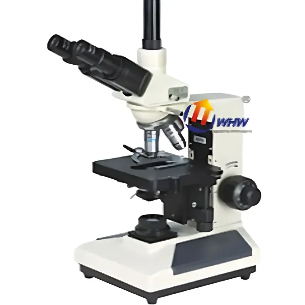
| Optical Configuration | Trinocular Head (30° Inclined, Interpupillary Adjustment 55–75 mm) |
| Eyepieces | Widefield WF10×/18 mm (Standard), Optional: WF16×, WF20×, P16× |
| Objective Lenses | Achromatic 4×, 10×, 40× (spring-loaded), 100× (spring-loaded, oil immersion) |
| Mechanical Stage | Dual-layer, 160 mm × 140 mm platform, 80 mm × 50 mm travel range |
| Condenser | NA 1.25 Abbe condenser with adjustable iris diaphragm and filter holder |
| Focus Mechanism | Coaxial coarse/fine focus |
| fine focus graduation | 2 µm per division |
| Illumination | 6 V / 20 W halogen lamp, AC 220 V / 110 V compatible, brightness continuously adjustable, Köhler illumination capable |
| Optional Accessories | Darkfield & phase contrast kits, teaching head, mirror, camera adapters (for DSLR/CCD), image measurement software, semi-plan and plan apochromatic objective sets (20×, 60×, oil-immersion variants) |
Overview
The YANRUN XSP-8CA Trinocular Biological Microscope is an upright, research-grade optical instrument engineered for routine and advanced life science applications in academic laboratories, clinical pathology units, and quality control environments. Designed around classical brightfield microscopy principles, it employs a robust, thermally stable metal frame and precision-ground achromatic optics to deliver high-fidelity, low-aberration imaging across the visible spectrum (400–700 nm). Its trinocular optical path—featuring a 30° inclined head with interpupillary adjustment (55–75 mm)—enables simultaneous visual observation and high-resolution digital documentation via a dedicated photo port. The microscope adheres to international optical design conventions defined in ISO 8578:2017 (Microscopes — Requirements for biological microscopes) and supports Köhler illumination alignment, ensuring uniform field illumination and optimal contrast-to-noise ratio for unstained and stained biological specimens.
Key Features
- Trinocular head with 30° inclination and ergonomic interpupillary adjustment (55–75 mm), optimized for extended user comfort and dual-path imaging.
- Standard widefield eyepieces (WF10×/18 mm) provide a broad field of view and ample eye relief; optional high-magnification or compensating eyepieces (WF16×, WF20×, P16×) extend analytical capability.
- Achromatic objective suite (4×, 10×, 40×s, 100×s oil) delivers consistent color correction and flatness across the field, with spring-loaded front lenses safeguarding against slide contact during high-magnification operation.
- Dual-layer mechanical stage (160 mm × 140 mm) offers precise, backlash-free X–Y translation (80 mm × 50 mm range) with vernier scales for repeatable specimen repositioning—critical for serial section analysis or multi-location imaging protocols.
- NA 1.25 Abbe condenser with iris diaphragm and filter holder enables full numerical aperture utilization and contrast modulation; compatible with optional darkfield and phase contrast modules for enhanced visualization of transparent, unstained cells.
- Coaxial coarse/fine focusing system with 2 µm fine-focus graduation ensures sub-micron Z-axis repeatability—essential for z-stack acquisition and morphometric quantification.
- 6 V / 20 W halogen illumination system features continuous brightness control and built-in Köhler alignment capability, supporting standardized light management per ISO 10934-1 and ASTM E2822 guidelines.
Sample Compatibility & Compliance
The XSP-8CA accommodates standard 1 mm-thick glass microscope slides (76 mm × 26 mm) and circular coverslips (18 mm, 22 mm, 24 mm diameters). It is validated for use with common histological preparations (H&E, Giemsa, PAS), live-cell cultures in Petri dishes or chambered coverslips (with appropriate working distance objectives), and microbiological smears—including Gram-stained bacterial isolates. All optical components comply with ISO 8578:2017 mechanical and optical tolerances. The illumination system meets IEC 61000-6-3 EMC emission standards. When integrated with FDA 21 CFR Part 11–compliant image capture software (e.g., third-party GLP-certified packages), the system supports audit-trail-enabled documentation for regulated QC/QA workflows under GMP and CLIA frameworks.
Software & Data Management
The microscope’s trinocular port is mechanically and optically calibrated for seamless integration with C-mount or F-mount digital cameras (DSLR, CMOS, CCD). Standard adapter options include 1×, 0.5×, and 0.35× reduction lenses to match sensor size and preserve native resolution. Compatible image acquisition platforms support TIFF, PNG, and multi-layer TIFF export, with embedded metadata (objective magnification, exposure time, illumination intensity). Optional YANRUN-branded image measurement software provides calibrated length/area/volume quantification, particle counting, and histogram-based thresholding—all traceable to NIST-calibrated stage micrometers. Exported datasets conform to MIAME-compliant metadata schemas for downstream integration into LIMS or ELN systems.
Applications
- Hematology and clinical cytology: differential white blood cell counts, reticulocyte analysis, and platelet morphology assessment.
- Microbiology: Gram staining evaluation, fungal hyphae characterization, and motility assays using wet-mount preparations.
- Botanical and zoological education: vascular tissue sectioning, pollen grain morphology, and protozoan behavior studies.
- Pharmaceutical QC: excipient particle sizing, tablet coating uniformity inspection, and dissolution test residue analysis.
- Academic research: immunofluorescence co-localization (when paired with appropriate filter cubes), mitotic index scoring, and morphometric profiling of cultured cell lines (e.g., NIH/3T3, HeLa).
FAQ
Is the XSP-8CA suitable for oil immersion work?
Yes—the 100× objective is spring-loaded and designed for use with Type A immersion oil (nD = 1.515); proper cleaning protocol post-use is required to maintain lens integrity.
Can this microscope be used for phase contrast imaging?
Yes—phase contrast capability is available as an optional upgrade kit, including annular diaphragms and phase objectives (10×, 40×, 100×), compliant with ISO 8578 Annex B specifications.
What is the maximum usable magnification with standard eyepieces and objectives?
With WF10× eyepieces and the 100× oil objective, total magnification is 1000×; resolution limit is ~0.2 µm under ideal Köhler illumination and optimal sample preparation.
Does the illumination system support long-term stability during time-lapse imaging?
The halogen lamp exhibits minimal thermal drift over 60-minute intervals; for extended sessions (>2 hr), external LED illumination modules (sold separately) are recommended to reduce heat load on live specimens.
Are calibration certificates available for metrological traceability?
Yes—NIST-traceable stage micrometer and eyepiece graticule calibration kits are available upon request, supporting ISO/IEC 17025-aligned internal validation procedures.


