YANRUN BID-300 Inverted Biological Microscope
| Brand | YANRUN |
|---|---|
| Origin | Shanghai, China |
| Manufacturer Type | Direct Manufacturer |
| Product Category | Domestic (China-Made) |
| Model | BID-300 Inverted Biological Microscope |
| Instrument Type | Inverted Microscope |
| Eyepiece Configuration | Binocular |
| Optical Magnification Range | 100×–400× |
| Objective Conjugate Distance | Infinity |
| Eyepiece Port Diameter | 30 mm |
| Parfocal Distance | 10 mm |
| Eyepieces | Wide-Field WF10X (Φ22 mm), Centering Telescope |
| Infinity-Corrected Long Working Distance Achromatic Objectives (Cover Glass Thickness | 1.2 mm): 10X/0.25 (WD 19.4 mm), 20X/0.40 (WD 8.0 mm), 40X/0.60 (WD 3.5 mm) |
| Infinity-Corrected Phase Contrast Objectives (PHP) | 10X/0.25 (WD 4.3 mm) |
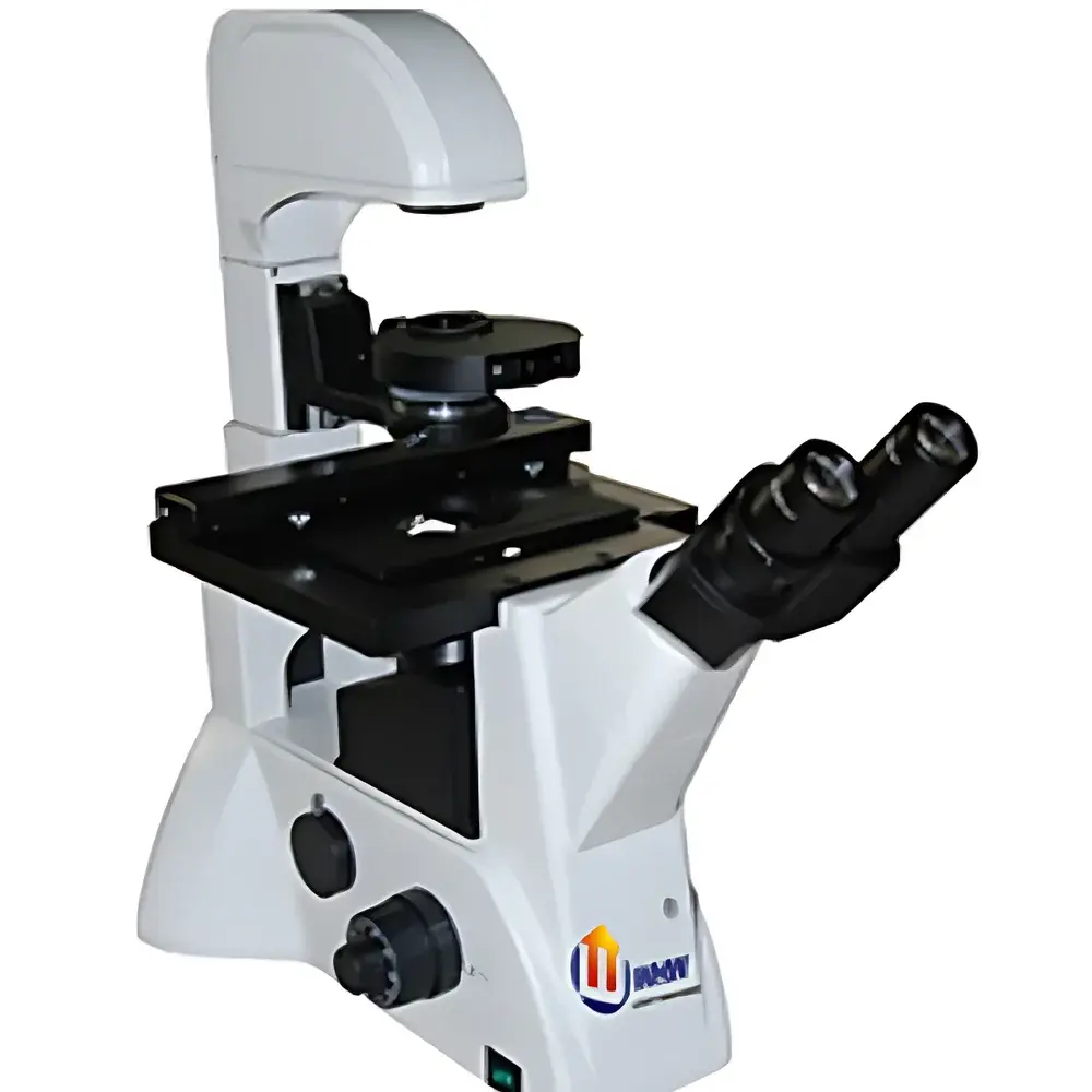
| Eyepiece Tube | Trinocular, 45° Inclination, Interpupillary Adjustment 53–75 mm |
| Focus Mechanism | Coaxial Coarse/Fine Focus with Adjustable Coarse Tension, Locking Mechanism, Limit Stop, Fine Focus Graduation: 2 µm |
| Nosepiece | Quintuple (5-Position) |
| Stage | Dual-Layer Mechanical Stage (227 × 208 mm), Travel Range: 134.5 × 77 mm, Removable Vernier Scale |
| Stage Plate | Circular Glass Plate (Φ118 mm) |
| Culture Dish Adapters | Three Interchangeable Plates — Type I (86 × 129.5 mm, fits Φ87.5 mm dish), Type II (34 × 77.5 mm, fits Φ68.5 mm dish), Type III (57 × 82 mm) |
| Condenser | Long Working Distance Condenser (WD 55 mm) with Rotating Phase Contrast Annulus Disc |
| Illumination | 6 V / 30 W Halogen Lamp, Adjustable Brightness and Height |
| Filters | Blue, Green, Ground Glass |
| Optional Accessories | Graticule Eyepiece (10X, 0.1 mm/div), Additional Phase Contrast Objectives (20X/0.40 PHP2, 40X/0.65 PHP2), Multi-Format Sample Holders (for 35 mm, 50 mm, T25, 66 × 66 mm square, and Standard Slide), Yellow Filter, Extended WD Condenser (WD 70 mm), Rotating Phase Contrast Disc |
Overview
The YANRUN BID-300 Inverted Biological Microscope is engineered for routine and advanced live-cell observation in academic research laboratories, biotechnology QC environments, and pharmaceutical cell culture facilities. Its inverted optical architecture positions the objective lenses beneath the specimen stage—enabling direct visualization of adherent or suspension cells within standard Petri dishes, multi-well plates, and cylindrical culture vessels without sample inversion or mounting. The system employs infinity-corrected achromatic and phase contrast optics, ensuring high-fidelity image formation across the full magnification range (100× to 400×) while maintaining parfocality and minimal chromatic aberration. Designed around Köhler illumination principles, the integrated 6 V / 30 W halogen light source delivers stable, adjustable intensity with spatially optimized condenser alignment—critical for quantitative phase contrast imaging and low-light digital acquisition.
Key Features
- Hinged trinocular head inclined at 45°, supporting simultaneous visual observation and high-efficiency photomicrography with 100% light path transmission—no beam-splitting loss.
- Dual-layer mechanical stage (227 × 208 mm) with 134.5 × 77 mm travel range and removable vernier scale; precision-engineered for reproducible XY positioning during time-lapse experiments.
- Three interchangeable culture dish adapters accommodate common vessel formats: Φ87.5 mm round dishes (Type I), Φ68.5 mm dishes (Type II), and rectangular configurations up to 57 × 82 mm (Type III), minimizing stage reconfiguration downtime.
- Long working distance condenser (55 mm) with rotating phase contrast annulus disc enables rapid switching between brightfield and multiple phase contrast modes (e.g., PH1, PH2, PH3)—compatible with cover glass thicknesses of 1.2 mm per ISO 8578.
- Coaxial coarse/fine focus mechanism with adjustable coarse tension, mechanical lock, upper/lower limit stops, and 2 µm fine focus graduation ensures stable Z-axis control during extended imaging sessions.
- Five-position nosepiece accepts industry-standard RMS-threaded objectives; includes pre-aligned infinity-corrected long WD achromats (10X/0.25, 20X/0.40, 40X/0.60) and dedicated phase contrast objectives (10X/0.25 PHP).
Sample Compatibility & Compliance
The BID-300 accommodates a broad spectrum of live biological specimens—including monolayer cultures (e.g., HEK293, HeLa, NIH/3T3), organoids in Matrigel® domes, spheroids in U-bottom plates, and suspension cells in chambered coverslips. Its 55 mm condenser working distance and modular stage adapters permit unobstructed access to tall vessels such as Erlenmeyer flasks and roller bottles. All optical components comply with ISO 10934-1 (Microscopes — Nomenclature of parts) and ISO 8578 (Microscopes — Requirements for phase contrast systems). While not certified for GMP manufacturing environments, the instrument supports GLP-compliant documentation workflows when paired with validated camera systems and audit-trail-enabled acquisition software.
Software & Data Management
The BID-300 features a rear-mounted C-mount (1×) interface compatible with industry-standard CMOS/CCD cameras (e.g., Sony IMX series, ON Semiconductor PYTHON sensors). When integrated with third-party imaging platforms—such as NIS-Elements (Nikon), ZEN (Zeiss), or open-source Micro-Manager—the microscope supports time-lapse acquisition, multi-channel fluorescence registration (with optional filter cubes), and metadata embedding (objective ID, magnification, exposure time, stage coordinates). Image files are exported in TIFF or OME-TIFF format, ensuring compatibility with FIJI/ImageJ, QuPath, and MATLAB-based analysis pipelines. Firmware-independent hardware triggering enables synchronization with external devices (e.g., environmental chambers, micromanipulators) via TTL signals.
Applications
- Real-time monitoring of cell proliferation, migration, and confluence in routine passage workflows.
- Phase contrast assessment of unstained primary neurons, stem cell differentiation, and epithelial-mesenchymal transition (EMT) morphology.
- Quality control of bioreactor harvests and cryopreserved vials prior to thawing and expansion.
- Pre-screening of transfection efficiency and CRISPR editing outcomes before downstream genotyping.
- Educational demonstration of mitotic stages, cytokinesis, and organelle dynamics in undergraduate life science laboratories.
FAQ
What is the maximum working distance available for objective lenses on the BID-300?
Standard included objectives provide working distances ranging from 3.5 mm (40X) to 19.4 mm (10X); optional extended WD objectives (e.g., 20X/0.40 PHP2, WD 8.0 mm) and a 70 mm condenser are available for thick-vessel applications.
Is the microscope compatible with fluorescence imaging?
The base configuration supports brightfield and phase contrast only; fluorescence capability requires optional filter cubes, excitation/emission filters, and a dedicated epi-illuminator—not included in standard delivery.
Can the BID-300 be integrated into automated incubator systems?
Yes—the mechanical stage and focus drive accept external motorization kits (e.g., Prior ProScan III, Ludl MAC5000), and the C-mount interface supports synchronized triggering with environmental controllers via TTL I/O.
Does the system meet FDA 21 CFR Part 11 requirements?
The microscope itself is not a regulated medical device; however, when used with validated, Part 11-compliant acquisition software and electronic lab notebooks (ELNs), it may support compliant data capture in regulated bioprocessing contexts.
What is the warranty coverage for the BID-300?
YANRUN provides a 24-month limited warranty covering defects in materials and workmanship under normal laboratory use; optical components are covered for 12 months against coating delamination or decentering.


