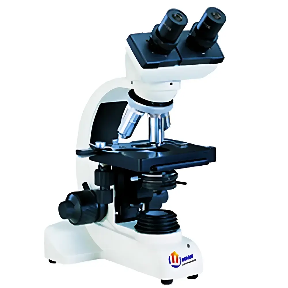
YANRUN BI-14 Biological Inverted Microscope
| Brand | YANRUN |
|---|---|
| Origin | Shanghai, China |
| Manufacturer Type | Direct Manufacturer |
| Country of Origin | China |
| Model | BI-14 Biological Microscope |
| Microscope Type | Inverted Microscope |
| Eyepiece Configuration | Binocular |
| Magnification Range (Standard) | 40×–1000× |
| Max Optional Magnification | 64×–1600× |
| Eyepieces | Widefield WF10× (Φ18 mm) |
| Objectives | Achromatic 4×/0.10, 10×/0.25, 40×/0.65 (spring-loaded), 100×/1.25 (spring-loaded, oil immersion) |
| Nosepiece | Four-position inward-rolling ball bearing turret with external positioning |
| Stage | Dual-layer mechanical stage (125 mm × 115 mm), travel range 70 mm × 30 mm |
| Condenser | Abbe condenser with adjustable iris diaphragm and filter holder, NA 1.25, vertically adjustable |
| Filters | Blue filter, ground glass |
| Illuminator | 6 V / 15 W halogen lamp with brightness control |
| Fine Focus Increment | 4 µm |
| Focus Mechanism | Coaxial coarse/fine focusing (BI-14A: standard |
| BI-14B | with limit lock, tension adjustment, and locking mechanism) |
| Tube Angle | 30° inclined, sliding binocular head |
| Optional Accessories | WF16× eyepieces, reticle eyepieces, monocular tube (45°), three-position turret, rotary or fixed stage variants, green/yellow filters, 5 W fluorescent lamp (110/220 V), high-intensity white LED illuminator, plano-concave mirror with stand, incandescent lamp, LED power adapters (9 V / 500 mA), USB/VIDEO trinocular photo-port, simple polarizing system (360° rotatable polarizer + push-pull analyzer) |
Overview
The YANRUN BI-14 Biological Inverted Microscope is engineered for routine and advanced life science applications requiring stable observation of living cells, tissue cultures, and adherent specimens in Petri dishes, flasks, and multi-well plates. Its inverted optical configuration places the objectives beneath the specimen stage and the condenser above—enabling unobstructed access to samples during manipulation, microinjection, or long-term time-lapse imaging. The microscope employs Köhler illumination principles via its NA 1.25 Abbe condenser and precision-halogen light source, ensuring uniform brightness, optimal contrast, and minimal thermal load on live specimens. Designed around a rigid cast-aluminum chassis and coaxial focusing architecture, the BI-14 delivers mechanical stability essential for reproducible quantitative microscopy and compatibility with auxiliary digital imaging systems.
Key Features
- Widefield optical path with achromatic objective lens set (4×/0.10, 10×/0.25, 40×/0.65 spring-loaded, 100×/1.25 oil immersion) delivering high chromatic correction and flat-field performance across the full magnification range (40×–1000× standard; extendable to 1600× with optional WF16× eyepieces)
- Binocular inclined viewing tube (30° tilt, sliding interpupillary adjustment) with widefield WF10× (Φ18 mm) eyepieces for extended user comfort and reduced eye strain during prolonged observation sessions
- Coaxial coarse/fine focusing mechanism with 4 µm fine-focus graduation—BI-14B variant includes dual-function lockable limit stop and adjustable coarse-drive tension for precise parfocal consistency and vibration resistance
- Dual-layer mechanical stage (125 mm × 115 mm) with calibrated X–Y translation (70 mm × 30 mm travel), compatible with standard culture vessels and supporting repeatable coordinate-based sample repositioning
- Abbe condenser (NA 1.25) with independently adjustable iris diaphragm and integrated filter holder—enables critical aperture control for optimizing resolution, depth of field, and contrast in both brightfield and phase contrast modalities
- 6 V / 15 W halogen illumination system with continuous dimming control and heat-absorbing filter integration, minimizing phototoxicity and thermal drift during live-cell observation
Sample Compatibility & Compliance
The BI-14 accommodates standard biological sample formats including 35 mm Petri dishes, T-25/T-75 flasks, 6–96-well plates, and custom chambered coverslips. Its low-profile stage design and extended working distance objectives support micromanipulation workflows without optical interference. While not certified to ISO 13485 or FDA 21 CFR Part 11 out-of-the-box, the instrument’s mechanical repeatability, stable illumination output, and standardized optical interfaces (e.g., trinocular port for camera coupling) align with GLP-compliant documentation practices when integrated into validated laboratory workflows. Optional polarizing and fluorescence accessories permit basic birefringence analysis and narrow-band epifluorescence screening—though dedicated fluorescence applications require externally filtered excitation sources and emission detection pathways.
Software & Data Management
The BI-14 supports third-party digital imaging via its trinocular photo-port (USB/VIDEO output), enabling direct integration with industry-standard acquisition platforms such as NIS-Elements (Nikon), ZEN (Zeiss), or open-source tools like MicroManager and Fiji/ImageJ. No proprietary firmware or closed software ecosystem is embedded—ensuring full audit trail transparency, raw data export (TIFF, BMP, AVI), and compatibility with LIMS or ELN systems. When paired with calibrated stage encoders or motorized focus modules (available as aftermarket upgrades), the platform meets baseline requirements for semi-automated image capture protocols referenced in ASTM E2811 and ISO/IEC 17025-accredited laboratories.
Applications
- Cell culture monitoring: Confluence assessment, morphology evaluation, and passage timing in primary and immortalized mammalian lines
- In vitro toxicology assays: Real-time observation of cytotoxic responses, membrane integrity changes, and organelle dynamics
- Embryology and developmental biology: Blastocyst staging, zygote cleavage tracking, and organoid growth characterization
- Microbiology: Motility analysis of protozoa and flagellated bacteria under low-magnification scanning
- Quality control in biomanufacturing: Sterility verification, mycoplasma screening, and adventitious agent detection per USP
- Educational laboratories: Foundational training in microscopy technique, staining interpretation, and optical parameter optimization
FAQ
Is the BI-14 suitable for oil immersion imaging?
Yes—the 100×/1.25 NA objective is specifically designed for immersion oil use and achieves theoretical resolution limits near 0.35 µm under optimal Köhler alignment.
Can the microscope be upgraded for fluorescence imaging?
It supports basic epifluorescence via optional 5 W fluorescent lamp and filter sets; however, high-sensitivity fluorescence requires external LED or mercury arc excitation sources with bandpass-filtered dichroics and emission optics.
What stage options are available for specialized sample holders?
Rotary (Φ120 mm), fixed (120 mm × 120 mm), and single-lens fixed-stage configurations are offered—each accommodating custom inserts for microfluidic chips or electrophysiology rigs.
Does the BI-14 meet regulatory requirements for GMP environments?
As a Class I general-purpose microscope, it provides the mechanical reliability and interface standards expected in GMP settings when deployed with documented calibration, preventive maintenance logs, and operator training records—but does not carry CE IVD or FDA 510(k) clearance.
How is illumination stability ensured over extended acquisition periods?
The regulated halogen power supply and integrated heat-absorbing filter maintain consistent color temperature and intensity for >4 hours at mid-range brightness settings, minimizing photobleaching and thermal drift in time-lapse experiments.


