Orient KOJI i-NIR-II Time-Resolved Small Animal In Vivo Fluorescence Imaging System
| Brand | Orient KOJI |
|---|---|
| Origin | Tianjin, China |
| Manufacturer Type | Authorized Distributor |
| Country of Origin | China |
| Model | i-NIR-II |
| Instrument Category | Optical Imaging System |
| Spectral Detection Range | 400–1650 nm |
| Spatial Resolution | 1 µm |
| Field of View | 75 mm × 75 mm |
| Temporal Resolution | 100 ps – 100 ms |
| Scan Resolution | 4096 × 4096 pixels |
| Scan Speed | 5 kHz |
| Maximum Penetration Depth | 5 mm |
| Excitation Laser Options | 785 nm, 808 nm, 980 nm, 1064 nm, 1310 nm (femtosecond pulsed lasers) |
| Sample Capacity | Single mouse (anesthetized, temperature- and respiration-controlled staging) |
Overview
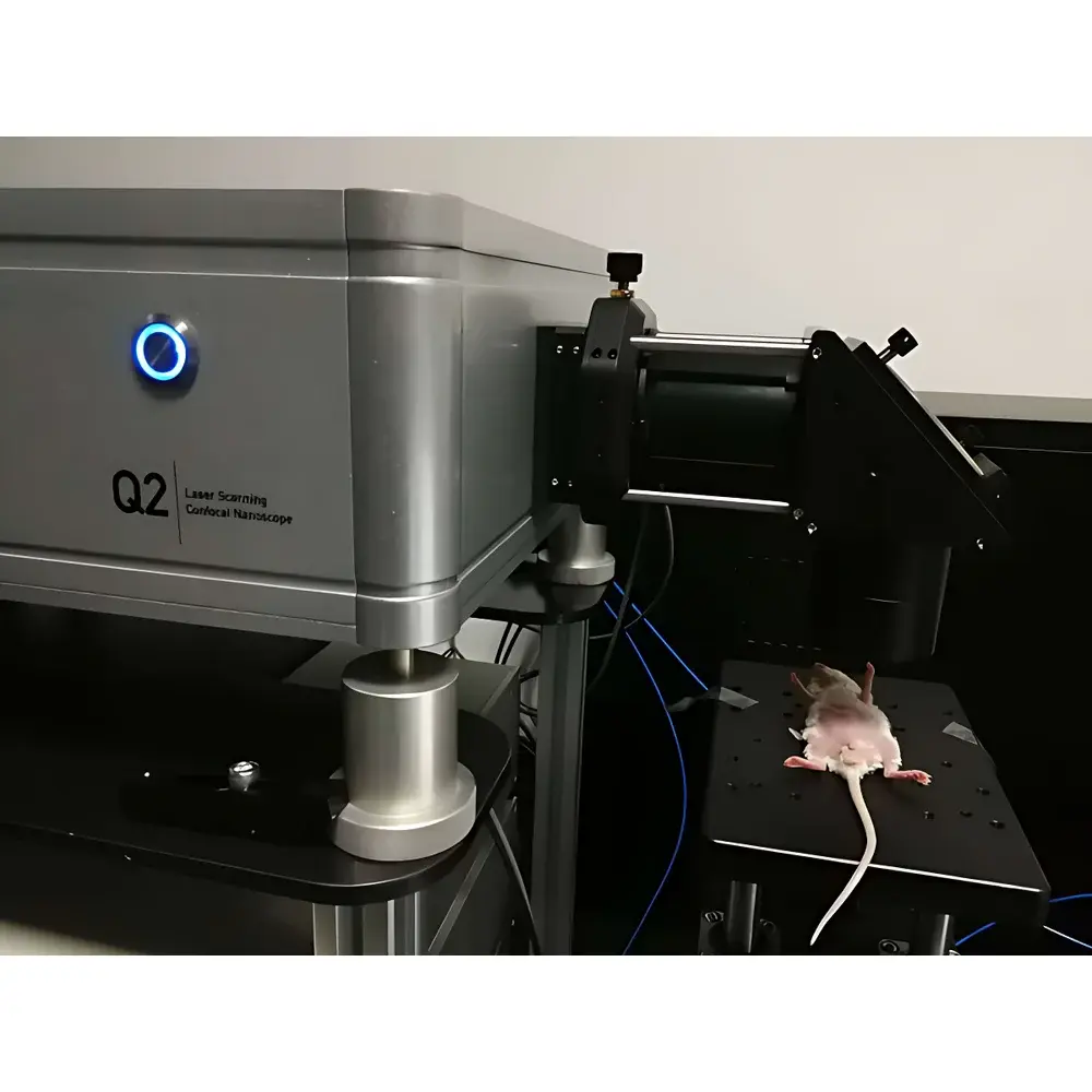
The Orient KOJI i-NIR-II is a high-performance, time-resolved small animal in vivo fluorescence imaging system engineered for quantitative, multi-parametric optical characterization in preclinical research. It operates on the principle of laser-scanning time-domain fluorescence lifetime imaging (FLIM) combined with spectral-domain intensity mapping, enabling simultaneous acquisition of fluorescence intensity, lifetime decay kinetics (τ), and spectral emission profiles across the visible to short-wave infrared (NIR-II) spectrum. Unlike conventional widefield or CCD-based systems, the i-NIR-II employs confocal point-scanning architecture with galvanometric mirrors and ultrafast pulsed excitation (femtosecond lasers), delivering diffraction-limited spatial resolution (≤1 µm) and picosecond-scale temporal fidelity. Its extended spectral sensitivity—spanning 400–1700 nm—supports dual-band operation: standard NIR-I (700–900 nm) and biologically advantageous NIR-II (1000–1700 nm), where reduced tissue scattering and autofluorescence significantly enhance signal-to-background ratio and imaging depth (up to 5 mm in murine tissue). The system is designed for longitudinal, non-invasive monitoring of molecular probes—including quantum dots, rare-earth-doped nanoparticles, and organic fluorophores—with intrinsic or environment-sensitive lifetimes.
Key Features
- Laser-scanning confocal architecture with galvanometric mirror positioning for precise pixel-by-pixel excitation and detection
- Dual-detector configuration supporting switchable spectral bands: NIR-I (400–1050 nm) and NIR-II (900–1700 nm), optimized via InGaAs or extended-range Si detectors
- Time-correlated single-photon counting (TCSPC) module enabling fluorescence lifetime quantification from 100 ps to 100 ms with <15 ps instrument response function (IRF)
- Adjustable field-of-view (FOV) ranging continuously from 3 mm × 3 mm (high-magnification cellular interrogation) to 75 mm × 75 mm (whole-body murine imaging)
- Integrated animal handling stage with real-time physiological monitoring (body temperature, respiration rate, O₂ saturation) and isoflurane anesthesia delivery
- Multi-wavelength femtosecond laser excitation platform (785 nm, 808 nm, 980 nm, 1064 nm, 1310 nm) with pulse width <150 fs and repetition rate ≥80 MHz
- 4096 × 4096 pixel scan matrix with sub-micron lateral sampling density and calibrated photometric linearity across five orders of magnitude
Sample Compatibility & Compliance
The i-NIR-II is validated for use with anesthetized C57BL/6, BALB/c, and nude mice (typically 20–30 g), accommodating standard dorsal/lateral/supine positioning. Sample preparation adheres to IACUC-compliant protocols; thermal regulation maintains core body temperature at 36.5 ± 0.5 °C throughout acquisition. The system supports quantitative comparison across longitudinal sessions through automated stage calibration and fiducial marker registration. Data output complies with MIAME/MINR guidelines for optical imaging metadata. Hardware and software architecture are compatible with GLP-aligned workflows: audit trails, user access controls, and electronic signatures meet requirements for pre-IND pharmacokinetic or biodistribution studies. While not FDA-cleared for clinical use, the platform conforms to ISO 13485–aligned manufacturing standards and electromagnetic compatibility per IEC 61326-1.
Software & Data Management
Acquisition and analysis are managed through the proprietary i-FLIM Suite v4.x, a Windows-based application built on Qt and HDF5 data architecture. The software provides real-time FLIM histogram fitting (multi-exponential decay models: mono-, bi-, tri-exponential; χ² minimization with Levenberg–Marquardt algorithm), lifetime color-mapping (τ-weighted pseudo-color LUTs), and spectral unmixing using constrained non-negative matrix factorization (cNMF). All raw TCSPC histograms, intensity stacks, and metadata are stored in vendor-neutral HDF5 files with embedded JSON sidecar descriptors. Export options include TIFF (16-bit), NIfTI-1 (for co-registration with MRI/PET), and CSV-compatible decay parameter tables. The suite supports 21 CFR Part 11–compliant configurations: role-based permissions, electronic signatures for report generation, and immutable audit logs covering acquisition parameters, user logins, and data export events.
Applications
- Quantitative pharmacokinetics and tumor-targeting efficiency of NIR-II contrast agents (e.g., Ag₂S QDs, PbS/CdS core-shell nanocrystals)
- Microenvironment sensing: pH-, ion-, or redox-dependent lifetime shifts in responsive probes within orthotopic tumor models
- Protein–protein interaction mapping via fluorescence resonance energy transfer (FRET) with lifetime-based ratiometric readout
- Angiogenesis and vascular permeability assessment using dynamic contrast-enhanced (DCE) FLIM protocols
- Neuroinflammation tracking with microglia-specific lifetime reporters in Alzheimer’s disease transgenic mice
- Therapeutic monitoring of photothermal or photodynamic therapy outcomes via lifetime quenching dynamics
FAQ
What is the minimum detectable lifetime shift that the i-NIR-II can resolve?
The system achieves a lifetime resolution of ≤25 ps under optimal signal conditions (SNR > 100:1), permitting discrimination of sub-nanosecond environmental perturbations in probe molecules.
Can the i-NIR-II perform simultaneous dual-channel FLIM with independent excitation wavelengths?
Yes—using time-gated detection and wavelength-selective dichroics, the system supports sequential or interleaved dual-excitation FLIM (e.g., 808 nm + 1310 nm) with cross-talk correction algorithms.
Is third-party probe validation supported?
Orient KOJI provides open API documentation and MATLAB/Python SDKs for custom probe calibration, including lifetime reference standards traceable to NIST SRM 2241 (fluorescein) and custom NIR-II lifetime phantoms.
How is photobleaching mitigated during long-duration scans?
The galvanometric scanning architecture enables dwell-time optimization and adaptive laser power modulation; average excitation fluence remains below 10 mJ/cm² per frame, well within ISO 60825-1 Class 1 safety limits for murine skin.
Does the system support co-registration with anatomical imaging modalities?
Yes—integrated fiducial markers and mechanical coordinate referencing allow rigid-body alignment with CT/MRI volumes; optional optical surface topography module enables deformable registration for soft-tissue warping correction.


