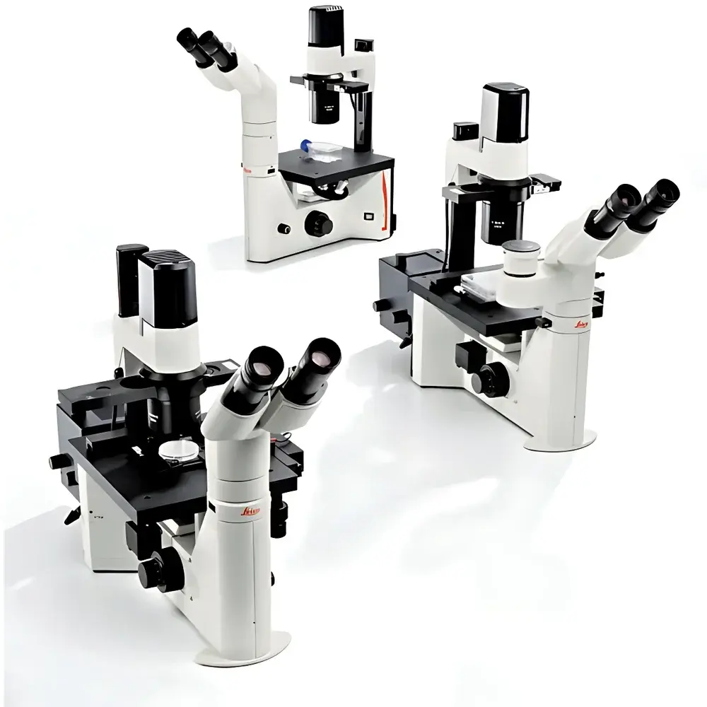
Leica DM IL LED Inverted Biological Microscope
| Brand | Leica |
|---|---|
| Origin | Germany |
| Model | DM IL LED |
| Instrument Type | Research-Grade Inverted Fluorescence Microscope |
| Excitation Source | Metal Halide Lamp (optional), LED-based Transmitted & Epi-Illumination |
| Medical Device Classification | Yes (CE-marked, Class IIa per MDR 2017/745) |
| Eyepiece Tube & Condenser | S40/0.45 and S80/0.30 |
| Objective | 40× Phase Contrast Objective (compatible up to 63×) |
| Fluorescence Module | 40 mm Filter Cube Mount |
| Light Source | 5 W High-Stability White LED (CCT ≈ 5,700 K) |
| Illumination Control | Fully Automated LED Brightness Regulation via Integrated Sensor Feedback |
| Focusing Mechanism | Coaxial Precision Focus Knobs with Fine/Coarse Adjustment |
| Condenser Working Distance | 200 mm (retractable design) |
| Observation Head | Trinocular, Ergonomic Adjustable Tube with Camera Port, DIC/Phase/IMC Compatibility |
| Stage | Heated Multi-Plate Cross-Stage (3-position, temperature-controlled to ±0.1 °C) |
| Fluorescence Filters | Standard 10× Widefield Fluorescence Set (BP 450–490 nm, BA 515 nm, LP 515 nm) |
Overview
The Leica DM IL LED is a research-grade inverted biological microscope engineered for high-fidelity live-cell imaging, long-term tissue culture observation, and integrated fluorescence applications in academic, pharmaceutical, and clinical research laboratories. Designed as Leica’s first dedicated inverted platform with fully integrated LED illumination, it employs a modular optical architecture grounded in HC (High Contrast) objective optics and Köhler-optimized transmitted-light pathways. Its core measurement principle relies on differential phase contrast modulation—specifically, both conventional phase contrast (PC) and Leica’s proprietary Integrated Modulation Contrast (IMC)—to generate label-free, high-contrast visualization of unstained, living specimens without introducing phototoxicity or thermal artifacts. The system supports simultaneous epi-fluorescence and transmitted-light observation, enabling correlative structural and functional analysis across modalities. All optical paths comply with ISO 10934-1 (Microscopy — Nomenclature of Components) and are calibrated to meet DIN EN 61000-6-3 (EMC immunity) and IEC 61000-6-4 (emission) standards for laboratory instrumentation.
Key Features
- Stable 5 W white LED illumination with 50,000-hour lifetime, constant color temperature (~5,700 K), and minimal radiant heat output—critical for thermosensitive live-cell assays.
- Dual condenser system: S40/0.45 (40 mm WD, NA 0.45) for high-resolution phase contrast at 5×–63×; S80/0.30 (80 mm WD, NA 0.30) for large-vessel compatibility (e.g., Petri dishes, bioreactors) with continuous height adjustment.
- Ergonomic trinocular head with variable tube height, interpupillary distance adjustment (55–75 mm), and diopter compensation (±5 dpt) to reduce operator fatigue during extended sessions.
- Heated 3-position cross-stage with independent temperature control (range: ambient to 40 °C, stability ±0.1 °C), supporting physiological conditions for stem cell differentiation, organoid development, and electrophysiology experiments.
- Automated illumination feedback loop: LED intensity dynamically adjusts when switching between brightfield, phase contrast, IMC, or fluorescence modes—ensuring consistent photon flux and eliminating manual recalibration.
- IMC slider-based modulation (no specialized objectives required) delivers pseudo-3D relief imaging comparable to DIC but with lower alignment sensitivity and broader compatibility across standard Leica HC PL FLUOTAR objectives (10×–40×).
Sample Compatibility & Compliance
The DM IL LED accommodates a wide range of specimen formats including glass-bottom dishes (35 mm, 60 mm), multi-well plates (6–96-well), perfusion chambers, and custom microfluidic devices. Its 200 mm working distance and unobstructed sample access enable integration with micromanipulators, patch-clamp rigs, and environmental control enclosures. As a CE-marked Class IIa medical device under EU MDR 2017/745, it meets essential requirements for safety and performance in diagnostic cytology (e.g., PAP smear evaluation) and in vitro diagnostic support. It conforms to ISO 13485:2016 quality management systems and supports GLP-compliant documentation through optional Leica Application Suite X (LAS X) audit trail logging (21 CFR Part 11 compliant when configured with user authentication and electronic signature modules).
Software & Data Management
The microscope operates natively with Leica Application Suite X (LAS X) Basic software, supporting real-time image acquisition, Z-stack reconstruction, time-lapse recording, and multi-channel fluorescence overlay. LAS X includes built-in calibration wizards for intensity normalization across illumination modes and automated focus mapping for stage drift correction. Raw data export follows TIFF 6.0 and OME-TIFF open standards, ensuring interoperability with ImageJ/Fiji, Python-based analysis pipelines (e.g., scikit-image, napari), and institutional LIMS platforms. Optional LAS X Life Science Edition adds AI-assisted cell segmentation, mitosis tracking, and quantitative fluorescence intensity profiling with background subtraction and bleed-through correction algorithms.
Applications
- Long-term live-cell imaging of adherent and suspension cultures under controlled thermal and gaseous environments.
- Phase contrast and IMC-based morphometric analysis of primary neurons, iPSC-derived cardiomyocytes, and tumor spheroids.
- Correlative brightfield/fluorescence workflows—for example, GFP-tagged protein localization overlaid on cytoskeletal structure visualized by IMC.
- Clinical cytology screening (e.g., cervical exfoliative samples) using standardized 10× fluorescence and high-NA phase contrast protocols aligned with CAP/CLIA guidelines.
- Electrophysiology support: low-heat LED illumination prevents local thermal perturbation of patch-clamp recordings; heated stage maintains membrane fluidity during prolonged whole-cell configuration.
- Developmental biology: time-lapse imaging of zebrafish embryos, C. elegans larval staging, and mouse blastocyst implantation assays.
FAQ
Is the DM IL LED compatible with third-party cameras and software?
Yes—via standard C-mount interface (1× magnification) and USB 3.0 or GigE Vision connectivity. SDKs for HALCON, OpenCV, and MATLAB are available upon request.
Can the S40 and S80 condensers be used interchangeably across all objectives?
Yes, both condensers support objectives from 5× to 63×; S40 is recommended for resolution-critical PC work at ≥40×, while S80 is optimized for large-volume culture vessels and IMC workflows requiring extended working distance.
Does the system support DIC (Differential Interference Contrast)?
No—DIC requires dedicated Nomarski prisms and strain-free objectives not included in the base configuration. IMC serves as a robust, objective-agnostic alternative for relief contrast in live-cell contexts.
What fluorescence excitation sources are supported beyond the standard LED module?
The DM IL LED Fluo variant accepts fiber-coupled EL6000 mercury-free excitation units and legacy Hg/Xe arc lamps via Leica’s universal fluorescence turret interface.
Is routine maintenance required for the LED illumination unit?
No scheduled maintenance is needed—the solid-state LED engine has no consumables, no alignment drift, and retains >95% luminance after 50,000 hours of operation.
Related Products


