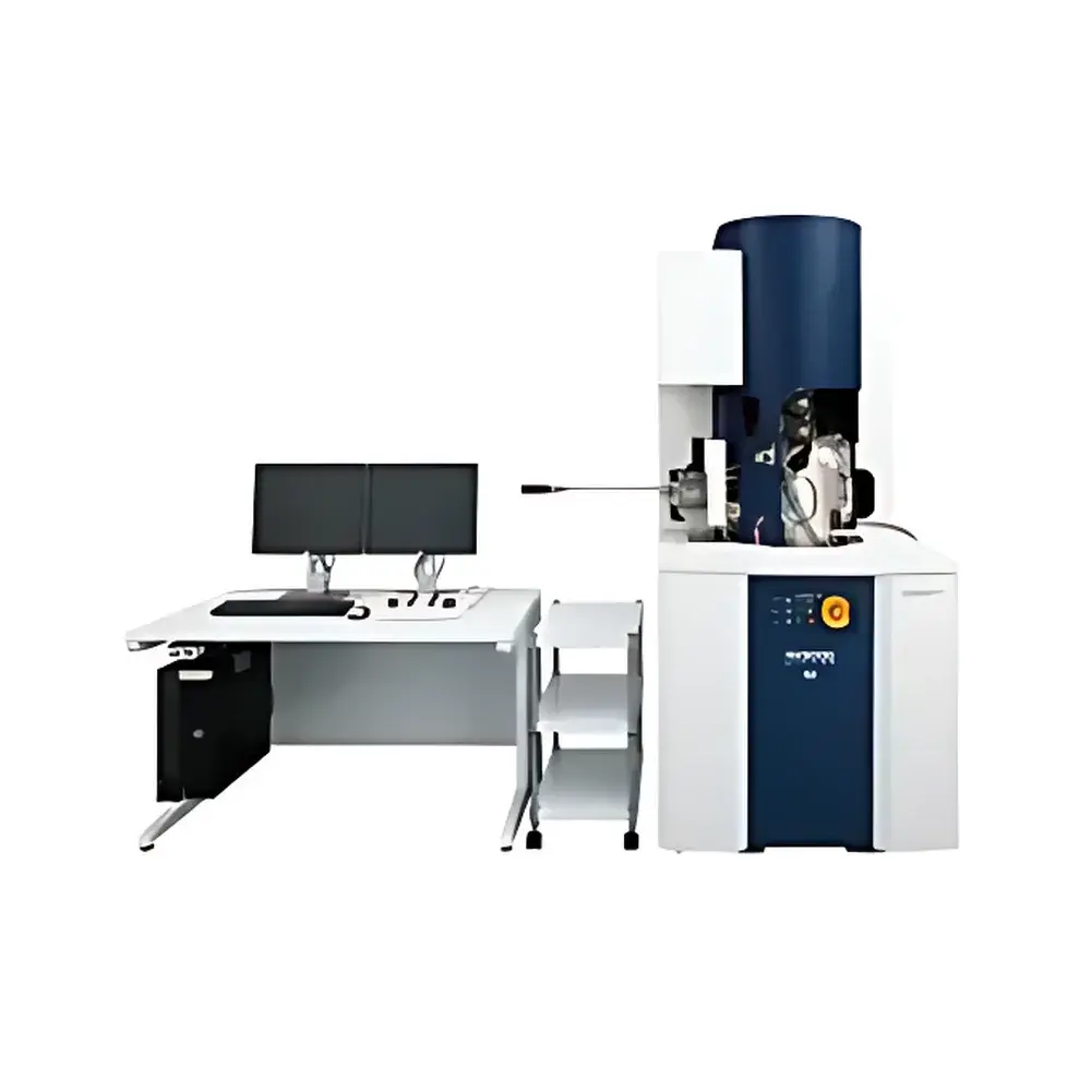
Hitachi NX9000 3D Analytical Focused Ion Beam-Scanning Electron Microscope System
| Brand | Hitachi |
|---|---|
| Origin | Japan |
| Manufacturer | Hitachi High-Tech Corporation |
| Type | Imported Instrument |
| Model | NX9000 |
| Pricing | Available Upon Request |
Overview
The Hitachi NX9000 is a high-precision, dual-beam analytical platform engineered for correlative 3D structural characterization at the nanoscale. It integrates a high-brightness cold field emission scanning electron microscope (CFEG-SEM) and a gallium liquid metal ion source focused ion beam (FIB) system in an orthogonal (90°) column architecture—optimized to eliminate geometric distortion inherent in tilted-axis FIB-SEM configurations. This orthogonal design enables true perpendicular incidence SEM imaging of FIB-milled cross-sections, ensuring faithful representation of native sample topography and microstructure without perspective warping or field-of-view drift during serial sectioning. The system supports automated, iterative cycles of site-specific ion milling, surface cleaning, and high-resolution SEM imaging—enabling robust acquisition of aligned, tomographic image stacks for volumetric reconstruction. Its architecture is purpose-built for quantitative 3D analysis across heterogeneous materials systems, from polycrystalline alloys and battery electrode architectures to hydrated biological tissues preserved via cryo-preparation.
Key Features
- Orthogonal dual-column configuration: SEM and FIB columns mounted at precisely 90°, enabling vertical-incidence SEM observation of horizontally milled surfaces—critical for artifact-free serial sectioning and registration accuracy.
- Cold field emission electron source (CFEG): Delivers stable, high-current-density electron beams with sub-nanometer probe size and exceptional signal-to-noise ratio for high-fidelity imaging of beam-sensitive and low-contrast specimens.
- High-sensitivity detection system: Includes in-lens secondary electron (SE), backscattered electron (BSE), and optional STEM-in-SEM detectors for multi-modal contrast acquisition during serial sectioning.
- Integrated Micro-sampling® module (optional): Enables precise, contamination-free extraction and transfer of lamellae for transmission electron microscopy (TEM) specimen preparation under controlled vacuum conditions.
- Triple Beam® capability (optional): Adds a second ion beam (e.g., Xe⁺ plasma FIB or O₂⁺ reactive beam) for accelerated milling, low-damage surface cleaning, or selective etching—extending analytical flexibility for hard, insulating, or multilayered samples.
- Automated serial sectioning workflow: Software-controlled synchronization of FIB milling depth, stage repositioning, and SEM image capture ensures sub-5 nm slice thickness repeatability and pixel-perfect stack alignment.
Sample Compatibility & Compliance
The NX9000 accommodates diverse specimen types—including conductive and non-conductive solids, cryo-fixed biological tissues, thin-film devices, and composite materials—without requiring extensive pre-treatment. Conductive coating is optional and minimized through low-voltage SEM imaging and charge compensation modes. For life science applications, the system supports cryo-transfer stages and in-chamber cryo-manipulation (when equipped with optional cryo-module), enabling structural preservation of neural tissue, organelles, and macromolecular assemblies. All hardware and software modules comply with ISO 14644-1 Class 5 cleanroom requirements for chamber integrity and are designed to meet GLP-compliant documentation standards. Data acquisition metadata—including beam parameters, stage coordinates, timestamp, and instrument configuration—is automatically embedded in image headers per ASTM E2821 and ISO/IEC 17025 traceability guidelines.
Software & Data Management
Hitachi’s proprietary 3D Analysis Suite provides end-to-end control—from region-of-interest designation and milling strategy definition to automated stack acquisition, drift correction, and volumetric reconstruction. Image registration employs feature-based affine and elastic algorithms to compensate for mechanical drift and thermal expansion over extended acquisition sessions (>24 h). Reconstructed volumes support quantitative morphometric analysis (e.g., pore network modeling, grain boundary mapping, synaptic density quantification) via export to standard formats (TIFF stack, HDF5, NIfTI). Audit trail functionality complies with FDA 21 CFR Part 11 requirements, logging all user actions, parameter changes, and calibration events with electronic signature support. Raw data and processed volumes are stored in vendor-neutral repositories compatible with institutional LIMS and FAIR data principles.
Applications
- Materials science: 3D grain boundary networks in Ni-based superalloys; porosity and crack propagation mapping in ceramic matrix composites; solid-electrolyte interphase (SEI) morphology in cycled Li-ion battery electrodes.
- Microelectronics: Through-silicon via (TSV) integrity assessment; interconnect delamination analysis; failure root-cause localization in advanced packaging.
- Life sciences: Nanoscale connectomics of murine hippocampal neurons; mitochondrial cristae architecture in cardiomyocytes; collagen fibril orientation in decellularized scaffolds.
- Geosciences: Pore-throat connectivity in shale matrix; mineral phase distribution in meteorite cross-sections; fluid inclusion geometry in metamorphic rocks.
FAQ
What is the typical slice thickness achievable with the NX9000 during serial sectioning?
Slice thickness is user-defined and typically ranges from 2 nm to 50 nm, depending on material hardness, beam current, and desired signal-to-noise ratio. Sub-5 nm slicing is routinely achieved on soft polymers and frozen-hydrated biological samples using low-kV FIB conditions.
Can the NX9000 perform in situ heating or electrical biasing during 3D analysis?
Yes—when equipped with compatible in situ holders (e.g., Protochips Fusion or DENSsolutions Climate), the system supports correlated structural evolution studies under controlled thermal or electrical stimuli, with real-time SEM/FIB feedback.
Is TEM lamella preparation supported natively within the NX9000 workflow?
Yes—the optional Micro-sampling® system enables fully integrated lift-out, thinning, and polishing of electron-transparent lamellae directly inside the chamber, minimizing handling-induced damage and contamination.
How does the orthogonal column design improve registration accuracy compared to conventional angled FIB-SEM systems?
By eliminating angular projection distortion and stage-tilt-induced parallax, the 90° geometry ensures each SEM frame captures the exact same physical plane milled by FIB—reducing lateral misregistration to 1,000-section stacks.


